Pt w advanced liver cirrhosis. 🏥 Comes w worsening ascites. No fever🤒, no bleeding🩸. 🧠 ok, no asterixis. BP 91/50. Labs📈: AKI (Cr 3.0 mg/dl), UNa 7 mEq/L, bland sediment. #POCUS 👉small cirrhotic liver with significant ascites. Paracentesis ruled out PBE. 📊Poll below👇 1/11
What would your initial treatment be? 2/11
Don't treat reflexively. A thorough physical exam ♥️🩺revealed a systolic murmur at left lower sternal border. Neck exam 👇

Sitting Down (90º) /// Supine (45º) 3/11
Albumin for volume expansion makes no sense in the face of venous congestion. Perform a good physical exam and you might not even need #POCUS. (Where is the fun in that? 😅)

If you look closely at the first video. IVC is huge! With no inspiratory variation. 4/11
#LUS (lung ultrasound) showed A pattern (not shown) which rules out lung congestion (good LV). The ♥️ is performing OK. 👇 PLAx, PSAx, A4ch with Good LV and RV systolic function. 5/11
#Echofirst revealed moderate/severe TR (but we knew this already, didn't we?). Apical 4ch, Parasternal Short and Parasternal Long views of the RV with color shown 👇 6/11
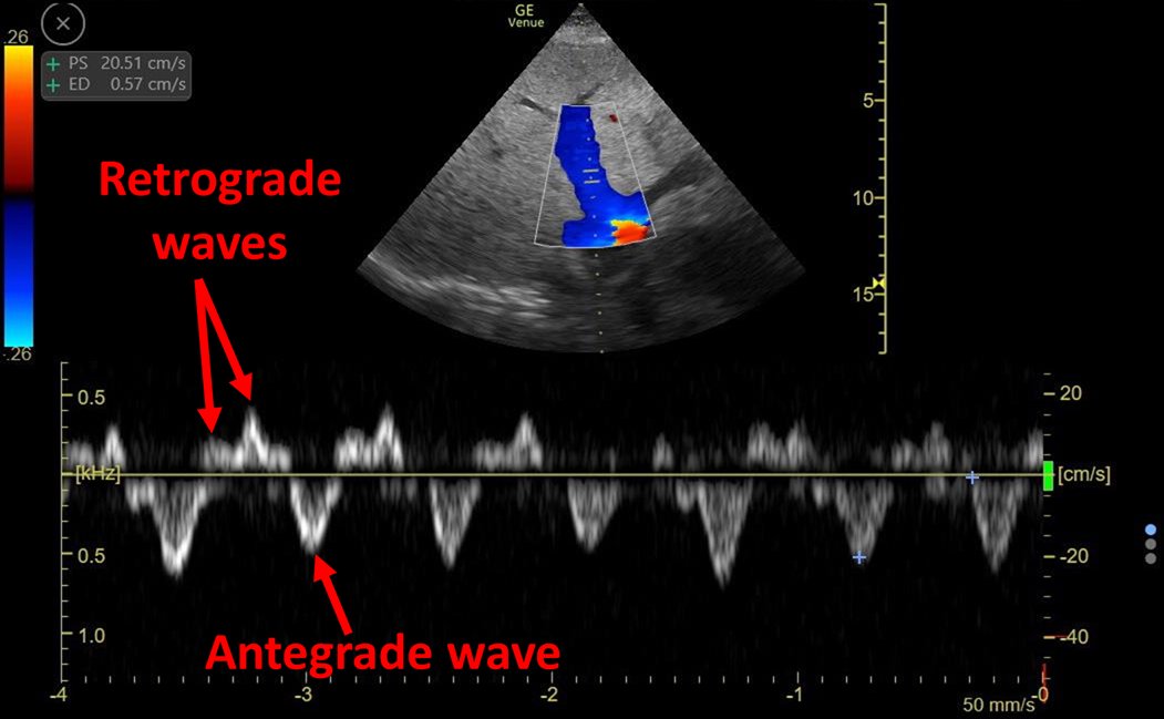
Some Doppler for the nerds: 1) TRVMax (showing moderate tricuspid regurgitation), 2) LVOT-VTI is increased (high stroke volume), 3) Normal shaped RVOT-VTI (ruling out severe precapillary PH) 4) Intra-renal venous doppler (IRVD) with biphasic pattern = venous congestion 7/11
#POCUS confirmed physical exam!

Hemodynamic AKI, distributive type.

Hyper-dynamic heart with increased CO (low peripheral vascular resistance) + Venous congestion = High output heart failure

8/11
Albumin and crystalloid are volume expanders. Why would we want this? This will make congestion worse. Remember volume expansion is meant to achieve an increase in cardiac output but... It is already very high!!

This quote here 👇👇👇👇

doi.org/10.1038/s41581… 9/11
In this case, I would advise to start vasopressors early (not wait 48 hrs of futile volume expansion). In fact, this was done and patient improved rapidly with good urine output and rapid resolution of AKI. 10/11
We see this frequently, with the exact same physiology: Low BP, normal ♥️, moderate/severe functional TR, and venous congestion (plethoric IVC and altered IRVD). I wrote about a very similar case here:

criticalcarenow.com/2020/10/04/alb…

11/11

• • •

Missing some Tweet in this thread? You can try to force a refresh
 

Keep Current with Eduardo R Argaiz

Eduardo R Argaiz Profile picture

Stay in touch and get notified when new unrolls are available from this author!

Read all threads

This Thread may be Removed Anytime!

PDF

Twitter may remove this content at anytime! Save it as PDF for later use!

Try unrolling a thread yourself!

how to unroll video
  1. Follow @ThreadReaderApp to mention us!

  2. From a Twitter thread mention us with a keyword "unroll"
@threadreaderapp unroll

Practice here first or read more on our help page!

More from @ArgaizR

10 Feb
📞 Nurse: Patient has a blood pressure of 226/118 mmHg!
📞 Resident: Nifedipine 30 mg STAT!
.....
📞 Me: About that last call, please hold Nifedipine until we assess the patient

A 🧵of some cases of Inpatient Hypertension 👇 1/15
🔎🖥️..This was a pt w ARDS on IMV. Other vitals 🆗. Previous BP= normal, no recent change in sedation, vent 🆗, no asynchronies. UOP = 0 for 2 hrs 🤔. Exam: Distended bladder!

After foley catheter change, BP normalised 😎

Why do we have this reflex to treat acute high BP? 2/15
A big component is the perceived expectation that we must do something! (In our minds, Are we trying to prevent organ injury?)

A great example can be seen here 👇 3/15

Read 15 tweets
16 Oct 20
Elderly ♂️, PMH of T2DM and CKD.

Suspected COVID-19 because of shortness of breath. Sent to the COVID-19 ED service.

No fever, no cough. No chest pain. Physical exam with patient sitting up (almost 90°):

¿Is this neck pulse arterial or venous? 1/8
Pulse is diffuse and the most striking feature is inward movement. I borrowed this table from @AndreMansoor's must-see lecture on Jugular Venous Pulse **Curiously, notice that there is a single peak instead of the expected double peak 🤔 2/8
I had to get my probe! #POCUS showed severely reduced EF with anterior wall motion abnormality and normal RV function.

EKG showed anterior ST segment changes. This was ACS! Cath lab was activated

3/8
Read 8 tweets
6 Oct 20
A case for hepatic vein Doppler lovers:

ECG tracing not abvailable on the ultrasound machine (I tried, I swear)

Can we still interpret this HV waveform without ECG?

I speculate we can!

I'll try to do it step by step: 1/7
What can we tell?

For one, there are 2 retrograde waves and 1 antegrade wave.

Given the patient was in sinus rhythm, then one of the retrograde waves MUST be an A wave! 2/7
Given HV waveform sequence should always be A-S-D, AND A wave is always retrograde:

Then this leaves only 2 possibilities: There could either be S wave reversal or D wave reversal 3/7
Read 8 tweets
5 Oct 20
Back on COVID-19 service this month

Went to see this patient with "increased respiratory drive despite high dose sedation and NMB". This is the vent: 1/4
Looking at the patient's monitor, the respiratory curve seems oddly coincident with heart rate: 2/4
Inspiratory pause reveals NO respiratory drive and several cardiogenic oscillations! 3/4
Read 4 tweets
14 May 20
🧵 AKI and #COVID19

68 yo ♂️ PMH obesity, HTN, CAD w stent, OSA, T2DM
➡️ ED w SOB + fever 39.9°C. Poor oral intake

RR 40, Sat 94% Room Air, BP 157/74 HR 124. Alert. Bibasilar crackles

Labs: Cr 1.3 (baseline 0.8), WBC 10, K 5.4, HCO3 17, CK 184. UA and CXR👇 (case from @NEJM)
How would you manage this AKI initially? What is the likely cause of AKI in patients with #COVID19? (this last question discussed in thread 🧵)
No easy answer except to say that FENa is very unlikely to be useful. It is not unreasonable to try fluids for AKI in the setting of perceived hypovolemia. However, this gets complicated when the potential for worsening ARDS exists. I'll try to tackle the answers one by one 💪
Read 17 tweets
11 May 20
Which of these patients has a more severe degree of venous congestion? #VExUS Thread 🧵 about the Portal Vein (1/17)
Video above shows IVC in short axis, long axis and diameter (from left to right)

Which of these patients has a more severe degree of venous congestion? (2/17)
Abdominal IVC size depends on the difference between CVP and IAP. At a constant IAP, IVC size will increase proportionally to CVP until it reaches the flat part of it's compliance curve. (Great thread by @Thind888 here: ) (3/17)
Read 18 tweets

Did Thread Reader help you today?

Support us! We are indie developers!


This site is made by just two indie developers on a laptop doing marketing, support and development! Read more about the story.

Become a Premium Member ($3/month or $30/year) and get exclusive features!

Become Premium

Too expensive? Make a small donation by buying us coffee ($5) or help with server cost ($10)

Donate via Paypal Become our Patreon

Thank you for your support!

Follow Us on Twitter!