1/
Hi #dermtwitter! I'm working with @jmervak et al, who put together this amazing #tweetorial on
NAILS!
Specifically, findings in the setting of systemic diseases: let nails help your diagnosis!
Coordinated with @naildisorders
#medstudenttwitter #medtwitter #meded #FOAMed
Hi #dermtwitter! I'm working with @jmervak et al, who put together this amazing #tweetorial on
NAILS!
Specifically, findings in the setting of systemic diseases: let nails help your diagnosis!
Coordinated with @naildisorders
#medstudenttwitter #medtwitter #meded #FOAMed

2/
First things first, do you mind telling us who you are?
First things first, do you mind telling us who you are?
3/Let's begin: Beau’s lines! Transverse depression across the nail. Means the nail briefly stopped growing and started again.
Seen weeks after nail injury! Or, if it's seen on multiple nails, ask about febrile illness (like post #covid19) or stressors like SJS or chemotherapy.
Seen weeks after nail injury! Or, if it's seen on multiple nails, ask about febrile illness (like post #covid19) or stressors like SJS or chemotherapy.


4/
If the injury is bad enough, the nail(s) will peel off, known as onychomadesis.
In kids a common cause of onychomadesis is:
If the injury is bad enough, the nail(s) will peel off, known as onychomadesis.
In kids a common cause of onychomadesis is:
5/
Correct answer is: Hand Foot Mouth Disease!!
Next up- Muehrcke’s lines! White transverse lines seen in chronic hypoalbuminemia such as nephrotic syndrome, liver disease or malnutrition.
Correct answer is: Hand Foot Mouth Disease!!
Next up- Muehrcke’s lines! White transverse lines seen in chronic hypoalbuminemia such as nephrotic syndrome, liver disease or malnutrition.

6/
But wait, did you know that the location of findings can clue you into different etiologies?!
Splinter hemorrhages closer to the lunula (white half moon at proximal end) are more closely correlated with SYSTEMIC DISEASE like endocarditis, while distally are likely TRAUMATIC.
But wait, did you know that the location of findings can clue you into different etiologies?!
Splinter hemorrhages closer to the lunula (white half moon at proximal end) are more closely correlated with SYSTEMIC DISEASE like endocarditis, while distally are likely TRAUMATIC.

7/
In addition to location, COLOR can tip you off too!
Proximal white subungual onychomycosis is a fungal infection type more commonly seen in those with HIV or other immunodeficiency. Seeing this should prompt testing for HIV.
In addition to location, COLOR can tip you off too!
Proximal white subungual onychomycosis is a fungal infection type more commonly seen in those with HIV or other immunodeficiency. Seeing this should prompt testing for HIV.

8/
Not all white discoloration of the nail is fungal! The differential for onycholysis ➜ or lifting of the nail, is long but includes thyroid disease, amyloidosis, and multiple myeloma.
Not all white discoloration of the nail is fungal! The differential for onycholysis ➜ or lifting of the nail, is long but includes thyroid disease, amyloidosis, and multiple myeloma.

9/
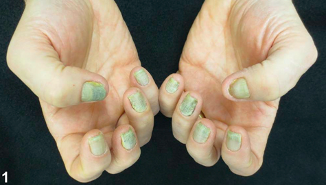
What if your whole nail is a different color.... like...Yellow!
Which specialty might need to see a yellow nail syndrome patient?
What if your whole nail is a different color.... like...Yellow!
Which specialty might need to see a yellow nail syndrome patient?
10/ PULMONOLOGY!
Yellow nail syndrome presents with slow nail growth, no lunula, lymphedema, and chronic respiratory disease such as bronchiectasis or pleural effusions. Nails can give you so much information about systemic processes! 👇👀
Yellow nail syndrome presents with slow nail growth, no lunula, lymphedema, and chronic respiratory disease such as bronchiectasis or pleural effusions. Nails can give you so much information about systemic processes! 👇👀
11/
Terry’s nails are seen in patients with cirrhosis, chronic renal failure, diabetes, and CHF. There is leukonychia (white) of the entire nail except for pink at at the tip. No lunula. However, they can also just be seen in normal aging!
#geriderm
Terry’s nails are seen in patients with cirrhosis, chronic renal failure, diabetes, and CHF. There is leukonychia (white) of the entire nail except for pink at at the tip. No lunula. However, they can also just be seen in normal aging!
#geriderm

12/
Lindsay’s nails are seen in chronic kidney disease! There is white proximally and reddish-brown color at the distal half of the nail.
Lindsay’s nails are seen in chronic kidney disease! There is white proximally and reddish-brown color at the distal half of the nail.

13/
Last one is for our fellow nervous Type A personalities!
Onychotillomania: nail picking
Onychophagia: nail biting
Last one is for our fellow nervous Type A personalities!
Onychotillomania: nail picking
Onychophagia: nail biting

14/ RECAP:
Nail exam findings can tell you about underlying systemic disease! #medicaldetective
Pay attention to characteristics like color, location, and orientation of findings-they give important clues about underlying pathology.
Nail exam findings can tell you about underlying systemic disease! #medicaldetective
Pay attention to characteristics like color, location, and orientation of findings-they give important clues about underlying pathology.
15/15
Thank you for joining us for @NailDisorders and @jmervak's first nail #tweetorial!
This was by no means a comprehensive list but using these observations can help lead to diagnosis (and make you look like a diagnostic rock star!)!
Thank you for joining us for @NailDisorders and @jmervak's first nail #tweetorial!
This was by no means a comprehensive list but using these observations can help lead to diagnosis (and make you look like a diagnostic rock star!)!
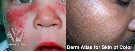
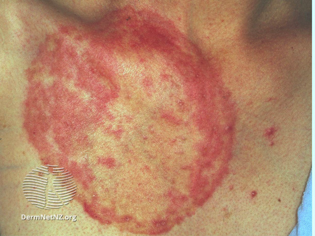
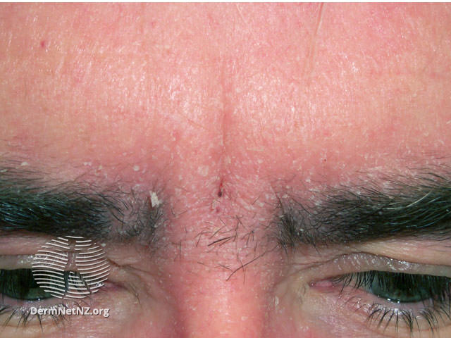
Examples of some of these nail disorders in darker skin tone👇👀
16/16
16/16
https://twitter.com/drsteventchen/status/1444703590543675404
• • •
Missing some Tweet in this thread? You can try to
force a refresh