1/
Let's spend some time on a #dermtwitter #tweetorial on all things....
HERPES!
Even though there are 8 herpesviruses that infect humans, I'm going to focus on HSV1 and HSV2 today.
#medtwitter #medstudenttwitter #meded #FOAMEd
Let's spend some time on a #dermtwitter #tweetorial on all things....
HERPES!
Even though there are 8 herpesviruses that infect humans, I'm going to focus on HSV1 and HSV2 today.
#medtwitter #medstudenttwitter #meded #FOAMEd

2/
You know how we say that everything could be sarcoid? Well, HSV-1/HSV-2 (which I'll refer to as herpes for this #thread) would be a close 2nd, ESPECIALLY on the inpatient service.
While HSV-1 is usually thought to be oral and HSV-2 genital, this certainly is NOT always true.
You know how we say that everything could be sarcoid? Well, HSV-1/HSV-2 (which I'll refer to as herpes for this #thread) would be a close 2nd, ESPECIALLY on the inpatient service.
While HSV-1 is usually thought to be oral and HSV-2 genital, this certainly is NOT always true.
3/
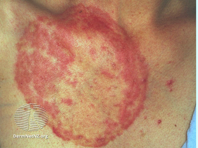
The class exam finding for herpes is the "dew drop on a rose petal." In clinical speak, that would be a vesicle on an erythematous base. But often we don't see the vesicle intact.
For ex, the photo above shows intact vesicles, whereas here, we just see the resultant crust.
The class exam finding for herpes is the "dew drop on a rose petal." In clinical speak, that would be a vesicle on an erythematous base. But often we don't see the vesicle intact.
For ex, the photo above shows intact vesicles, whereas here, we just see the resultant crust.

4/
So what does that mean? It means you have to have a high index of suspicion.
Any vesicle on a red base, or a small punched out ulcer surrounded with erythema could be herpes!
Often these vesicles join together to create a scalloped (bubbly) border!
So what does that mean? It means you have to have a high index of suspicion.
Any vesicle on a red base, or a small punched out ulcer surrounded with erythema could be herpes!
Often these vesicles join together to create a scalloped (bubbly) border!

5/
And furthermore, if a patient has an underlying rash (usually eczema), it can get superinfected with herpes, which we then call "eczema herpeticum."
This would look like monomorphic (similar looking) punched out ulcers on top of eczema! This is more common in kids!
And furthermore, if a patient has an underlying rash (usually eczema), it can get superinfected with herpes, which we then call "eczema herpeticum."
This would look like monomorphic (similar looking) punched out ulcers on top of eczema! This is more common in kids!

6/
Certain professions are also more prone to get subtypes of herpes. Herpetic whitlow is herpes (usually HSV-1) infection of the fingers.
Which profession do you think would predispose to this?
Certain professions are also more prone to get subtypes of herpes. Herpetic whitlow is herpes (usually HSV-1) infection of the fingers.
Which profession do you think would predispose to this?
7/
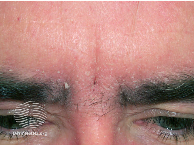
Dentists are ones at highest risk, which makes sense! Notice how the primary morphology is the same, just in a different place.
Clustered vesicles on an erythematous base with scalloped border--> just in a different body part!
Dentists are ones at highest risk, which makes sense! Notice how the primary morphology is the same, just in a different place.
Clustered vesicles on an erythematous base with scalloped border--> just in a different body part!

8/
TRANSMISSION: Usually from direct skin-skin or skin-infected fluid contact. That's why we see it transmitted between family, sexual partners, wrestlers, etc.
Interestingly, there's some evidence that HSV-1 seems to protect against HSV-2 infection, but not vice versa.🧐
TRANSMISSION: Usually from direct skin-skin or skin-infected fluid contact. That's why we see it transmitted between family, sexual partners, wrestlers, etc.
Interestingly, there's some evidence that HSV-1 seems to protect against HSV-2 infection, but not vice versa.🧐
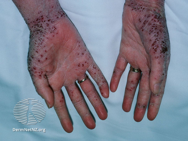
9/
But let's go back to why we have to always think about it. So many of my #dermtwitter colleagues and I have been fooled. Large ulcerations can be herpes (ESPECIALLY in the immunosuppressed)!
Always look for the scalloped border & erythematous rim. That's your usual giveaway.
But let's go back to why we have to always think about it. So many of my #dermtwitter colleagues and I have been fooled. Large ulcerations can be herpes (ESPECIALLY in the immunosuppressed)!
Always look for the scalloped border & erythematous rim. That's your usual giveaway.

10/
Don't forget HSV can infect so many other things than the skin. Keratitis, hepatitis, encephalitis, esophagitis, just to name a few.
Treatment is almost always one of the acyclovir antivirals. Valacyclovir is nice because it's dosed less frequently with good bioavailability.
Don't forget HSV can infect so many other things than the skin. Keratitis, hepatitis, encephalitis, esophagitis, just to name a few.
Treatment is almost always one of the acyclovir antivirals. Valacyclovir is nice because it's dosed less frequently with good bioavailability.
11/
Exception: In our cancer patients or immunosuppressed patients who get acyclovir prophylaxis, if you see a lack of response to therapy, consider sending a culture for sensitivities. These patients are more likely to have acyclovir resistant HSV, requiring the use of foscarnet
Exception: In our cancer patients or immunosuppressed patients who get acyclovir prophylaxis, if you see a lack of response to therapy, consider sending a culture for sensitivities. These patients are more likely to have acyclovir resistant HSV, requiring the use of foscarnet
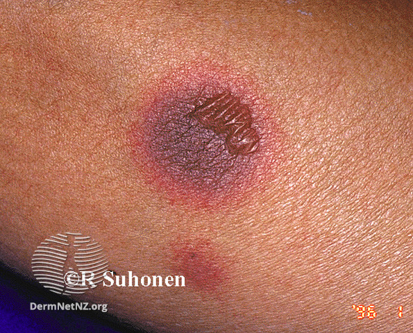
12/
One last #pearl for you all:
HSV is one of 2 main diagnoses in #dermatology where the lesion comes and goes in the EXACT same spot.
If you get that history, think of HSV (pic1) or fixed drug eruption (pic2), which thankfully are pretty easy to distinguish on exam.
One last #pearl for you all:
HSV is one of 2 main diagnoses in #dermatology where the lesion comes and goes in the EXACT same spot.
If you get that history, think of HSV (pic1) or fixed drug eruption (pic2), which thankfully are pretty easy to distinguish on exam.

12/
SUMMARY:
✅HSV1 and 2 are two of the herpesviruses
✅Vesicles on an pink base that cluster to create a scalloped border
✅ HSV can present atypically, esp with underlying conditions like eczema, immunosuppression
✅ Something that comes and goes in the same spot - think HSV.
SUMMARY:
✅HSV1 and 2 are two of the herpesviruses
✅Vesicles on an pink base that cluster to create a scalloped border
✅ HSV can present atypically, esp with underlying conditions like eczema, immunosuppression
✅ Something that comes and goes in the same spot - think HSV.
13/13
Thanks for joining for this whirlwind of a #tweetorial on #herpes! It can be tricky, but with a high index of suspicion, you'll never miss it again!
I didn't touch on a lot in an effort to keep it as high yield as possible, so feel free to add more below!
Until next time!
Thanks for joining for this whirlwind of a #tweetorial on #herpes! It can be tricky, but with a high index of suspicion, you'll never miss it again!
I didn't touch on a lot in an effort to keep it as high yield as possible, so feel free to add more below!
Until next time!
14/14
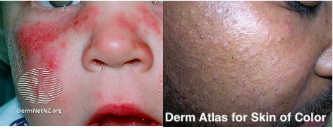
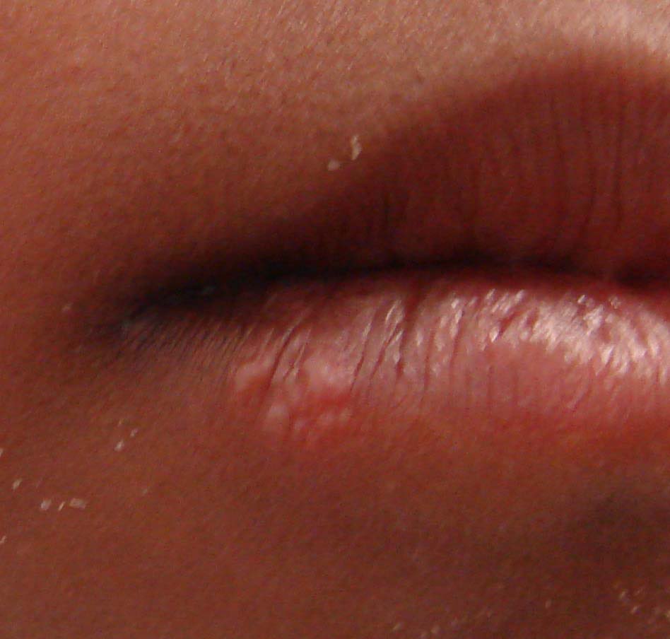
Oops, I lied. One more tweet to emphasize the importance of knowing how the exam differs in darker skin. While erythema is harder to see, the primary lesion is still notable as is the scalloped edge!
Pc: infectiousdiseaseadvisor.com/home/decision-…
Oops, I lied. One more tweet to emphasize the importance of knowing how the exam differs in darker skin. While erythema is harder to see, the primary lesion is still notable as is the scalloped edge!
Pc: infectiousdiseaseadvisor.com/home/decision-…
15/15
If you liked this type of stuff, consider checking out and following our new #dermatology #podcast, @TheDermConsult! 👇👇👇
I’m joined by @DrAveryLaChance and Bina Kassamali as we dissect mystery skin disease cases with you on our virtual consult team!
If you liked this type of stuff, consider checking out and following our new #dermatology #podcast, @TheDermConsult! 👇👇👇
I’m joined by @DrAveryLaChance and Bina Kassamali as we dissect mystery skin disease cases with you on our virtual consult team!
https://twitter.com/thedermconsult/status/1353320392660635648
• • •
Missing some Tweet in this thread? You can try to
force a refresh