link.springer.com/article/10.100…
Mini-Tweetorial Follows:
#CardioOnc #cvImaging #ACCimaging
@onco_cardiology @JStojanovskaMD @MichaelCoMD
@DmitryAbramovMD @chiarabd @HeartDocSubha @larsgrowo
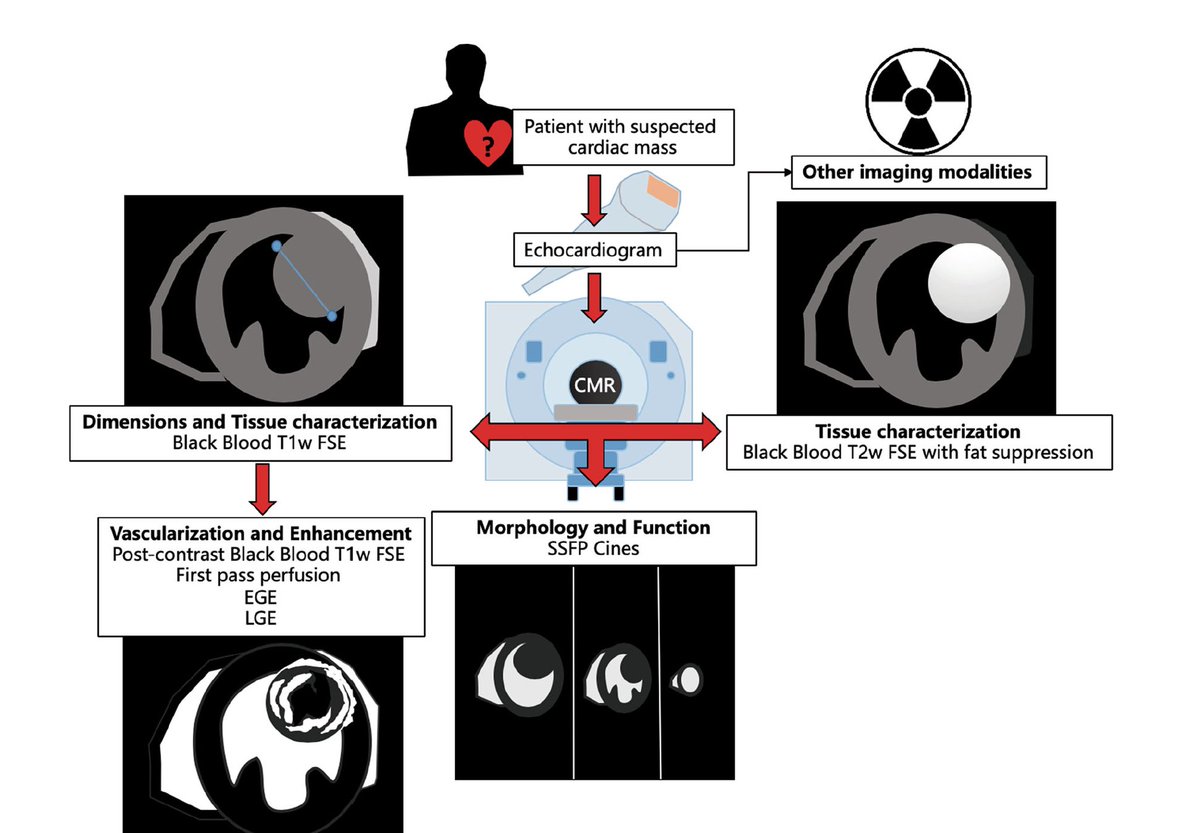
Primary Cardiac Tumors 0.056%
Can be benign, malignant or pseudotumor
Primary Cardiac tumor- mostly benign
#Cardiac Mass eval: detailed clinical presentation, History, Physical Exam, #echofirst to see the tumor characteristics
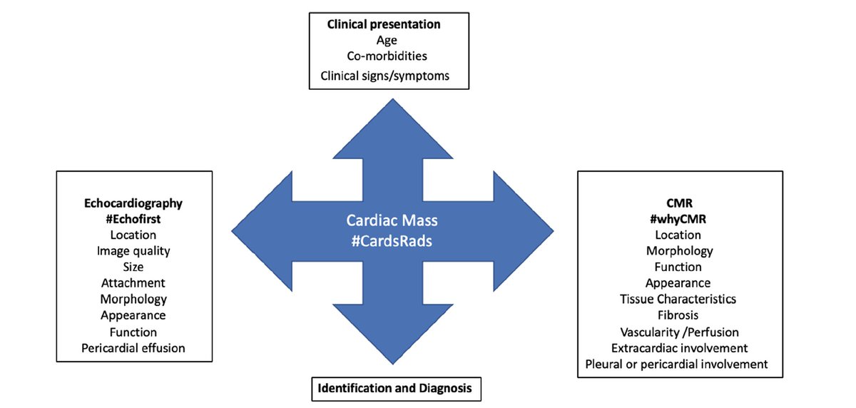
#whyCMR eval:
Cine imaging
T1 and T2 weight imaging with and without FS
First Pass Perfusion
EGE, LGE & post-Gad T1W imaging
@fabrizioricci @MasriAhmadMD @cshenoy3 @afshinfarzan
Myxoma:
Most frequent cardiac Tumor
commonly seen in the LA attached to the IAS
#whyCMR
T1W: Iso or Hypointense
T2W: High intensity
LGE: mostly Heterogenous enhancement
@Hragy @mirvatalasnag
2nd most common cardiac Tumor
75% of valvular tumor
#whyCMR
T1W: Isointense
T2W: Iso or Hypointense
LGE: near Homogenous enhancement
8.4% of all benign cardiac tumors
usually found in the LV, RA or IAS
#whyCMR
T1W and T2W: HYPER intense with suppression on Fat Sat prepulse
LGE: No enhancement
6% of all benign tumors.
most often seen in children and are rare in adults
multiple, small, lobulated, hyperechogenic and can be found intramyocardial
T1W: Iso-intense
T2W: Iso-intense to mildly hyperintense
LGE: No enhancement
more common in the pediatric population.
generally seen in isolation and mostly localized to left ventricle, can be seen in atria especially with polyposis syndrome
T1W: Iso-intense
T2W: Hypo-Intense
LGE: Hyper-Intense (Light-Bulb Enhancement)
5%-10% of all primary benign cardiac tumors
T1W: Iso-intense
T2W: Iso or Hyperintense
LGE: Heterogenous Uptake
Most common primary Malignant Tumor
Main role of #whyCMR is the extension into cardiac & extra-cardiac structures
Can have heterogenous appearance on #whyCMR
1.3% of all primary cardiac malignancies
T1W: Iso-intense to hypointense
T2W: Iso or mildly Hyperintense
LGE: Variable uptake
substantially more common than primary malignancy
T1-Wimaging of most secondary cardiac malignancies is low intensity & high signal intensity on T2-W imaging
A notable exception to this is metastatic melanoma, which appears hyperintense on T1-W imaging
The differential diagnosis can be challenging given the rarity of the tumors.
Thank you @onco_cardiology for this opportunity.
It was a fun project with one of the biggest #HeforShe in my professional life.


