This is an incredibly important preprint to inform #LongCovid. Among many analyses, the team recruited 4 patients w/ prolonged + recurrent olfactory function loss after #COVID-19 (time from first COVID-19 symptoms to inclusion ranged from 110-196 days): biorxiv.org/content/10.110…
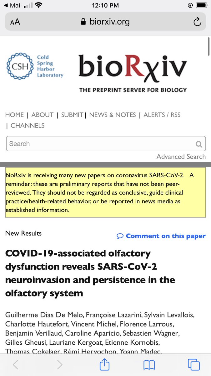
2/ None of these patients had detectable COVID-19 #RNA in nasopharyngeal samples by routine diagnosis (RT-qPCR). However, ALL patients had detectable COVID-19 RNA in samples obtained from their olfactory mucosa (confirmed with aRT-qPCR SYBR technique)
3/ Three of the patients had a high COVID-19 #viral load in the olfactory mucosa. Immunostaining additionally revealed the presence of COVID-19 antigens in 3 out of 4 patients. Based on that and related findings the team concluded...
4/ “...#SARS-CoV-2 has a significant tropism for the olfactory mucosa and, most importantly, we demonstrate that it can persist locally, not only a few weeks after general symptoms resolution but during several months in both mature and immature olfactory sensory #neurons.”
5/ In a further set of experiments, in addition to the olfactory bulb, #COVID-19 RNA was also detected in more remote #brain areas of infected hamsters, such as the cerebral cortex and the brainstem
6/ Leading the team to reference this separate Lancet study, which found COVID-19 viral RNA/protein in the brainstem of COVID-19 human patients via autopsy: thelancet.com/journals/laneu…
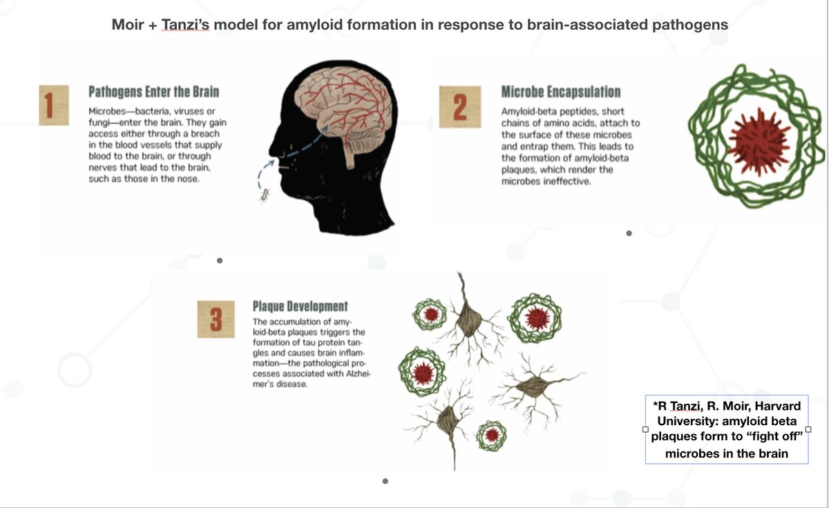
7/ I want to further note that the overall trend (certain #pathogens may infect the brain via the nose/olfactory bulb is central to Harvard’s late Rob Moir + Rudy Tanzi’s model for CNS pathogen activity in Alzheimer’s: pubmed.ncbi.nlm.nih.gov/30314800/
8/ Takeway: we are not going to understand #LongCovid (or #Alzheimer’s or #MECFS for that matter) unless we 1) analyze patient tissue/biopsies and use techniques that go far beyond routine testing 2) are open-minded to #pathogen entry + persistence in the central nervous system
• • •
Missing some Tweet in this thread? You can try to
force a refresh
1/ This important new study found that herpes simplex #virus 1 infection can drive the formation of tau tangle aggregates - protein clumps that facilitate the death of neurons in the #Alzheimer’s brain: nature.com/articles/s4159…
2/ The work was done by the same Harvard team who previously showed that the amyloid "plaque" in the Alzheimer's brain also forms in response to viral, bacterial, or fungal infections: pubmed.ncbi.nlm.nih.gov/30001512/
3/ Both findings suggest that Alzheimer's disease can be driven by persistent #infections in brain tissue. Amyloid plaques & tau tangles form IN RESPONSE to these infections as protective mechanisms the immune system has evolved to counter pathogen proliferation over time
Our new review is out! We detail mechanisms by which the viral, bacterial, parasite, and other pathogens that infect humans over a lifetime accelerate features of aging: sciencedirect.com/science/articl…
2/ Specifically, viruses such as the herpesviruses, as well as intracellular bacteria & parasites—express proteins and metabolites capable of interfering with host immune signaling, #mitochondrial function, gene expression, and the #epigenetic environment.
3/ Pathogen activity also contributes directly to age-related disease development: for example, #Alzheimer’s amyloid-β plaque can act as an antimicrobial peptide that forms in response to infection. pubmed.ncbi.nlm.nih.gov/30001512/
2/ Consider for example this study. The team identified dozens of viral proteins that distort human pathway signaling controlling #aging-associated processes such as senescence and apoptosis: pubmed.ncbi.nlm.nih.gov/36649176/
3/ Or this study - which found that human herpesvirus 6 can directly integrate into host telomeres. Telomeres carrying an integrated copy of the virus were shorter and more unstable: academic.oup.com/nar/article/42…
If you are a patient that meets #ME/CFS and #PEM criteria, info on the specific #infections or exposures that led to onset or exacerbation of your symptoms is of major importance. That info will help you pursue personalized treatment.
2/ That is because many of the infections you've sustained may still be #persisting in your body - in your tissue or nerves. These persistent infections can cause PEM symptoms by driving #mitochondrial dysfunction, blood vessel/perfusion issues, or #vagus nerve dysfunction
3/ If your symptoms started - or were exacerbated - by a #herpesvirus infection, such viruses persist in your system for life. Thus, treatment with herpesvirus #antivirals (e.g. the Pridgen Protocol which uses valacyclovir and Celebrex) has helped certain ME/CFS patients improve
Jawdropping data here showing dozens of #viruses - many rarely even discussed or tested for on a regular basis - in sewage collected from wastewater in multiple USA cities. The viruses are identified via unbiased sequencing that can identify any viral genome in the samples.
2/ Because the viruses are being identified in wastewater it's possible that some viruses are harbored by animals - for example cattle or birds - whose feces end up in the wastewater
3/ However, it's likely that most of the viruses being shed into wastewater come from infected humans. Viruses like the enteroviruses A, B, C, D68, Rhinoviruses A, B, C, Rotaviruses, Noroviruses, Rotaviruses, Mastadenoviruses, Adenoviruses, Rhinoviruses, Influenza viruses, etc.
Glad to have contributed to this new preprint. We found that some PVS participants had higher levels of circulating spike protein compared to controls.
This parallels #LongCovid where persistence of the SARS-CoV-2 #virus in patient tissue may also cause spike to periodically leak into blood
2/ For example, this study found #SARS-CoV-2 proteins including spike up to 1 year post-COVID in up to 25% of people tested. But identified spike was not a result of the COVID vaccine, since nearly all study participants had not received the vaccine: thelancet.com/journals/lanin…
3/ The same team also found SARS-CoV-2 spike protein encoding double-stranded RNA in LongCovid #gut tissue almost 2 years post-#infection. Such RNA is produced during active viral replication and thus wld not be vaccine derived: science.org/doi/10.1126/sc…