A 35-YO♂️: a 9-month of white concretions encircling the hairs in both axillae (A).

A Wood’s lamp🔎: yellow-green fluorescence of the concretions (B).

Dermoscopy: cottonlike structures on the hair shafts (C).
1/6

DOI: 10.1056/NEJMicm2206453
#dermatology #dermatología Image
TRICHOMYCOSIS AXILARIS

✔️ a superficial bacterial infection of the hair of the axillae
✔️misnomer, given that it is caused by corynebacterium species as opposed to fungi.
2/6

#dermtwitter #microbiology
After 1 week of treatment with topical clindamycin and a daily benzoyl peroxide wash, the patient’s symptoms abated and did not recur.
3/6

#MedTwitter #MedStudentTwitter
TRICHOMYCOSIS AXILARIS is characterised by yellow, black or red granular nodules or concretions that stick to the hair shaft.

It can also affect pubic hair (when it is called trichomycosis pubis), scrotal hair, and intergluteal hair.
4/6

#resident #MedicalStudents
Trichomycosis axillaris is caused by the overgrowth of Corynebacterium (Corynebacterium tenuis, C propinguum, C flavescens) and Serratia marcescens.

The concretions consist of tightly packed bacteria.
5/6

#bacteriology #IDtwitter
Visible deposits form on hair shafts when bacteria mix with dried apocrine sweat, especially in the presence of hyperhidrosis or impaired hygiene.
6/6

#Doctor #medicine #MedEd

• • •

Missing some Tweet in this thread? You can try to force a refresh
 

Keep Current with Antonio Guerrero

Antonio Guerrero Profile picture

Stay in touch and get notified when new unrolls are available from this author!

Read all threads

This Thread may be Removed Anytime!

PDF

Twitter may remove this content at anytime! Save it as PDF for later use!

Try unrolling a thread yourself!

how to unroll video
  1. Follow @ThreadReaderApp to mention us!

  2. From a Twitter thread mention us with a keyword "unroll"
@threadreaderapp unroll

Practice here first or read more on our help page!

More from @guerreroespejo

Dec 12
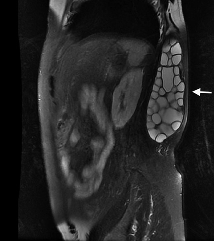
A 65-YO man, from North African: asymptomatic dorsolumbar mass for 30 years with normal skin appearance.

MRI: ?
1/6

doi.org/10.1093/cid/ci…
#radiology #MedTwitter #parasitology ImageImage
MRI: a multivesicular cyst in the latissimus dorsi muscle (arrow)

🧪for Echinococcus granulosus: ELISA, & Western blot➕
Surgery: multiple daughter vesicles of E. granulosus, with the presence of multiple scolices

INTRAMUSCULAR HYDATID CYST
2/6

#parasites #medicine #radres ImageImage
No puncture should be performed, to avoid dissemination of the cysts that can cause an anaphylactic shock.

The patient received 3 other courses of albendazole: serologic control became negative after 1 year.
3/6

#IDtwitter #surgery
Read 6 tweets
Dec 8
Lactante, 9 meses: lesiones peribucales, edema palpebral, conjuntivitis, xerosis peribucal con costras melicéricas, eritrodermia generalizada y lesiones en cuello, extremidades superiores y glúteos, con signo de Nikolsky +.
1/5

#pediatria #AtencionPrimaria #urgencias
SINDROME DE PIEL ESCALDADA ESTAFILOCÓCICA (Enfermedad de Ritter).

📌frecuente en < 5 años,
📌eritrodermia con signo de Nikolsky
📌conjuntivitis y edema facial con descamación periorificial.
2/5

continuum.aeped.es/screens/play/1…
@ContinuumAEP @aepediatria #infecciosas #microbología
@ContinuumAEP @aepediatria La enfermedad de Ritter está causada por cepas de estafilococo productoras de toxinas que se dirigen al complejo desmogleína 1 en la zona granulosa de la epidermis, provocando la exfoliación de la piel.
3/5

#MIR #MedTwitter
Read 5 tweets
Dec 5
A 59-YO DM, steroid, myasthenia gravis with thymoma: headache & deviation of tongue to the R side with fasciculation suggestive of right 12th cranial nerve palsy

CT: sphenoid sclerotic changes, soft tissue density lesion with calcification.
1/9

#neurology #radiology #IDtwitter Image
MRI brain: a hypointense lesion involving the body of sphenoid (R > L) and clivus on T2 weighted images and T1 weighted images with moderate contrast enhancement possibly fungal in aetiology.
2/9

doi.org/10.1016/j.idcr…
#radiologist #radres #MedTwitter ImageImage
Endoscopic sphenoidotomy & debridement: invasive fungal sphenoid sinusitis & CENTRAL SKULL BASE OSTEOMYELITIS involving the clivus

🔬from sphenoid mucosa: morphologically ASPERGILLUS with foci of tissue invasion.
3/9

#microbiology #maxillofacial #Orthopedics
Read 8 tweets
Dec 3
A 5-YO♂️ with a history of IgA vasculitis & a pustular nasal lesions had started scabbing after 2 days of systemic glucocorticoids: progressively painful skin ulcerations of L forearm (A), R calf (B) & R foot (C).
1/4

DOI: 10.1056/NEJMicm2204111
#Pediatrics #dermatology
Biopsy samples from all three lesions showed a dense neutrophilic infiltrate with negative tissue cultures.

PYODERMA GANGRENOSUM
2/4

#MedTwitter #Doctor #MedStudentTwitter
Laboratory & radiographic: no signs of underlying systemic disease, such as cancer, inflammatory bowel disease, or autoimmune conditions.

After 8 months of glucocorticoids: the lesions resolved completely.
¾

#resident #pediatric #MedicalStudents
Read 4 tweets
Dec 3
2022 #Monkeypox #Outbreak:
Confirmed cases, WHO, 02 December 2022

With the exception of West & Central Africa, the outbreak continues to primarily affect:
📌men who have sex with men
📌who have reported recent sex with one or multiple partners.
1/11

worldhealthorg.shinyapps.io/mpx_global/
WHO assesses the #Monkeypox global #Risk as Moderate.

Regionally, risk:
✔️as High in the Americas
✔️as Moderate in the African Region, Eastern Mediterranean Region, European Region & the South-East Asia Region.
✔️as Low in the Western Pacific Region

2/11
The 10 most affected countries:
📌United States of America
📌Brazil
📌Spain
📌France
📌Colombia
📌The United Kingdom
📌Germany
📌Peru
📌Mexico and
📌Canada

Together, these countries account for 86.0% of the #monkeypox cases reported globally.
3/11
Read 11 tweets
Nov 23
A 25-YO♂️ in Nigeria 2 years earlier: recurrent itchy swellings on his hands and forearms, appearing every week or so for the past few months.

What's in the #eye (A) & in the blood (B)?
1/4

doi.org/10.1016/S0140-…
#Ophthalmology #parasitology #microbiology ImageImage
Identification of:
📌an adult worm in the eye
📌the microfilariae on a blood smear

LOA LOA FILARIASIS is a skin and eye disease caused by the nematode worm Loa loa.
2/4

#parasites #MedEd #MedTwitter ImageImage
Loa loa is one of three parasitic filarial nematodes that cause subcutaneous filariasis in humans.
The other two are Mansonella streptocerca and Onchocerca volvulus (causes river blindness).
3/4

#Doctor #MedStudentTwitter #oftalmologia
Read 4 tweets

Did Thread Reader help you today?

Support us! We are indie developers!


This site is made by just two indie developers on a laptop doing marketing, support and development! Read more about the story.

Become a Premium Member ($3/month or $30/year) and get exclusive features!

Become Premium

Don't want to be a Premium member but still want to support us?

Make a small donation by buying us coffee ($5) or help with server cost ($10)

Donate via Paypal

Or Donate anonymously using crypto!

Ethereum

0xfe58350B80634f60Fa6Dc149a72b4DFbc17D341E copy

Bitcoin

3ATGMxNzCUFzxpMCHL5sWSt4DVtS8UqXpi copy

Thank you for your support!

Follow Us on Twitter!

:(