,
18 tweets,
5 min read
Read on Twitter
1/ New paper on “Quantifying how staining methods bias measurements of neuron morphologies”:
frontiersin.org/articles/10.33…
The logic of the paper:
frontiersin.org/articles/10.33…
The logic of the paper:
2/ The morphology of neurons has amazed neuroscientists from the dawn of neuroscience. Ramón y Cajal, the pioneering neuroscientist, drew many neurons using the Golgi staining method. 

3/ Since then many techniques have been developed to image neuron morphologies. These techniques involve the preparation of the specimen and the imaging.
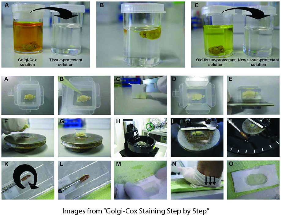
4/ One of the most important methodological choices is the staining method -- contrasting the neuron from the background. They help us to reconstruct the neuron.
5/ We test if "the same biological neurons collected with distinct staining methods have the same morphological (dendrites) features". In other words, do staining methods bias extracted morphologies?
6/ Here two neurons count as biologically similar if their rodent species, experimental condition, sex, age, brain region, and cell type are the same.
7/ To test this hypothesis, we search the public morphology repository, neuromorpho.org, to find comparison groups and identify 22 groups that match on biological features but differ on staining method.
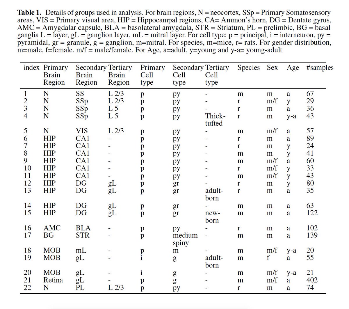
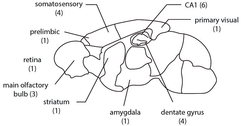
8/ The comparison groups cover many rodent brain regions.
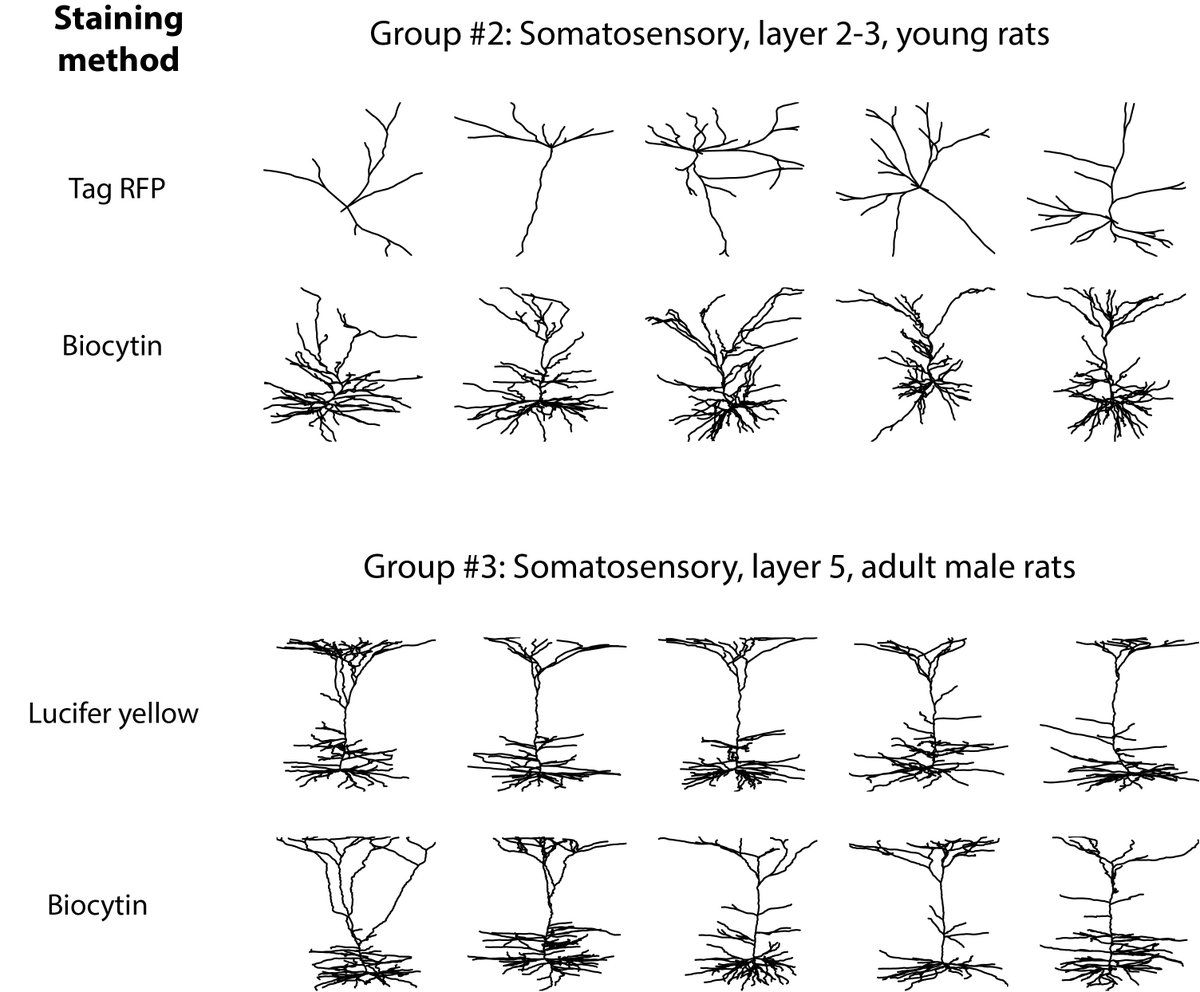
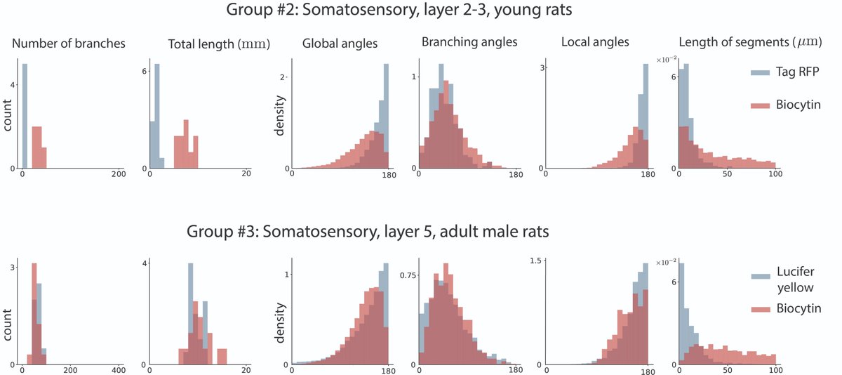
9/ For example, here are dendritic trees for two groups of somatosensory neurons labeled by two different staining methods
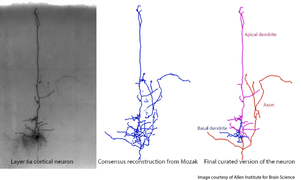
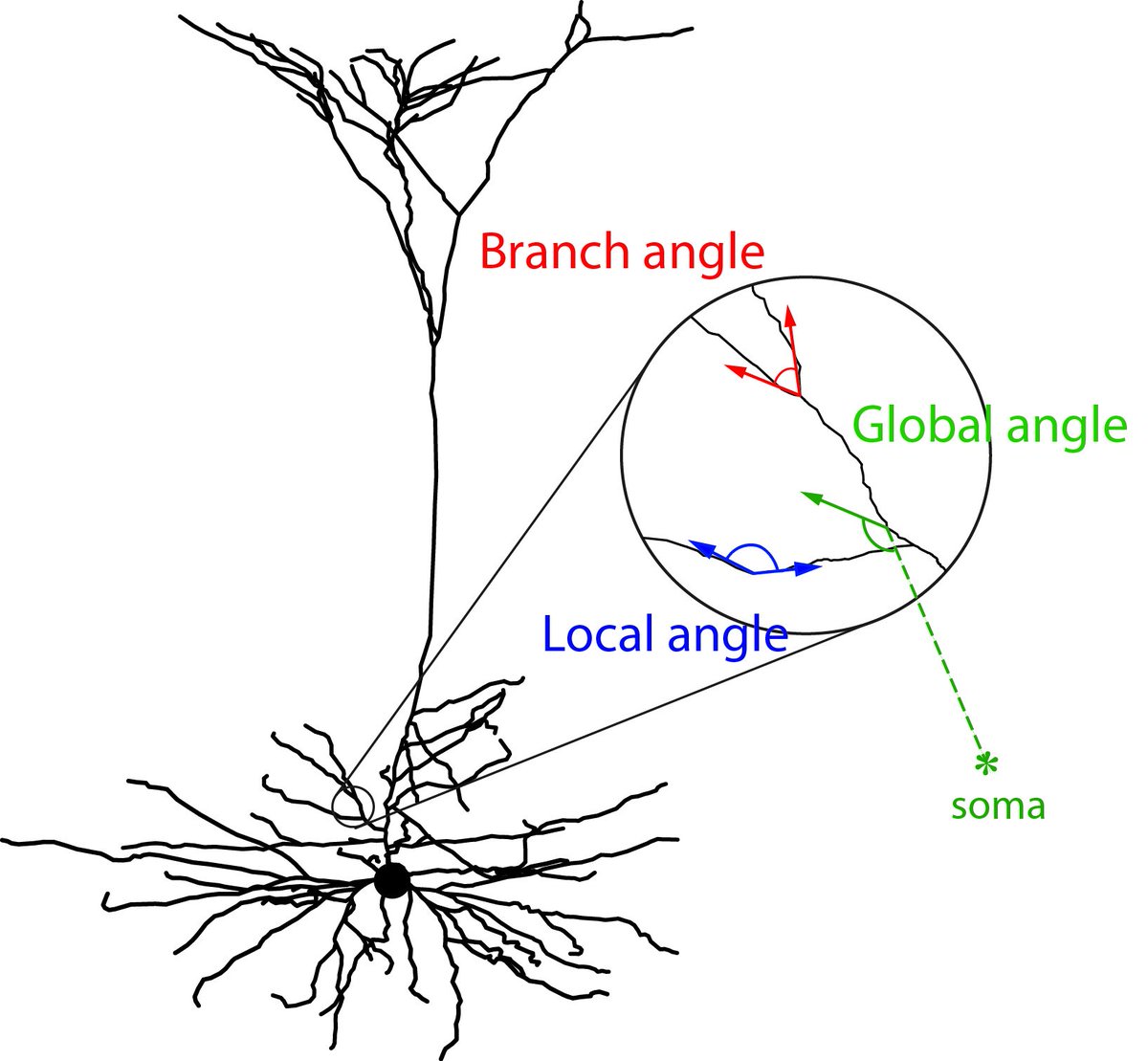
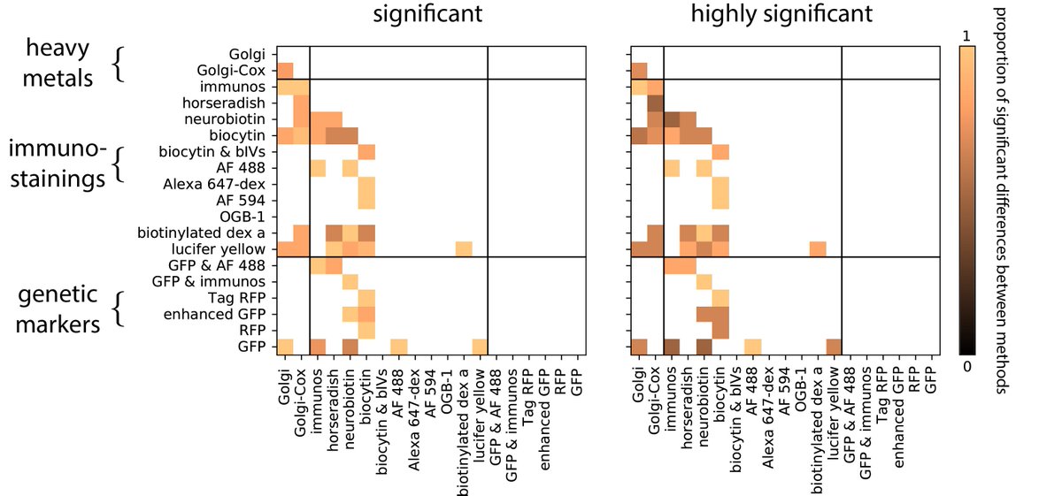
10/ To compare neurons extracted by different staining methods, we compute six morphological features that are sensitive to the local and global structure of the neuron.
11/ For these example groups, the histogram of these features shows large differences across many features.
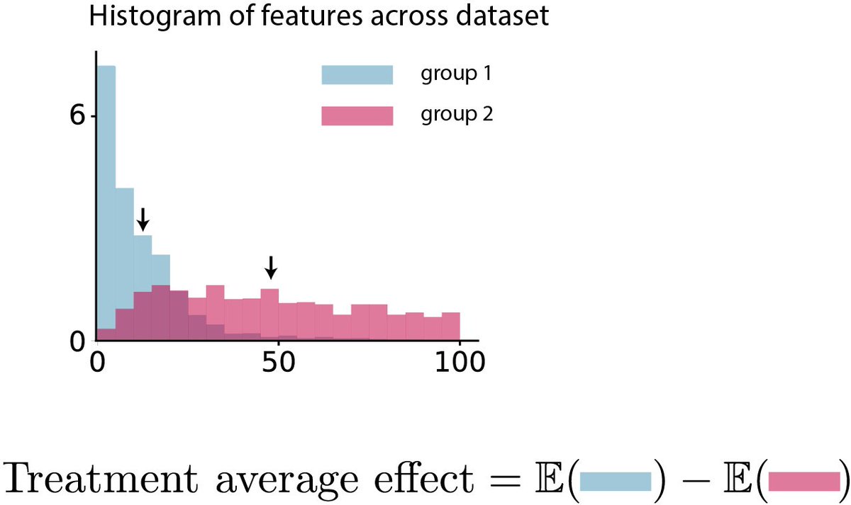
12/ How should we compare the features? For each of the 22 comparing groups and each feature, compute the average effect of the staining method on that feature.
13/ Applying the analysis for all pairs of staining methods (if a comparison group was available) rejects the hypothesis: Staining method change the morphology of the neuron massively.
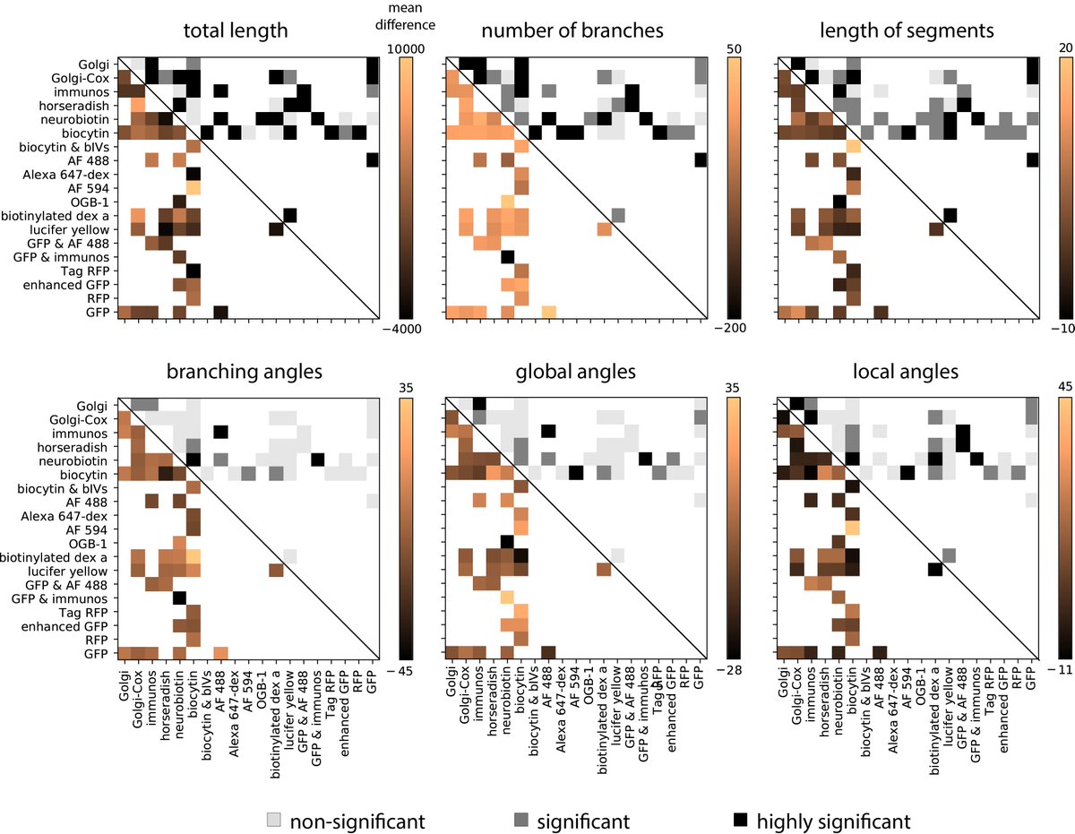
14/ Testing for a difference in any of the morphological features between two staining methods (Wilcoxon-rank sum test, details in the paper) produces similar results.
15/ Morphological data from many labs and brain regions are increasingly available. Our analysis tells a cautionary tale about studying neuron morphology when drawing from different datasets.
16/ However, we should be careful to jump to conclusions. For example, there may exist differences caused by details that happen to be correlated with the staining method, or current biological identification of neurons are not enough to define cell classes.
18/ This work would not have been possible without my amazing collaborators/mentors/friends @benlansdell @KordingLab