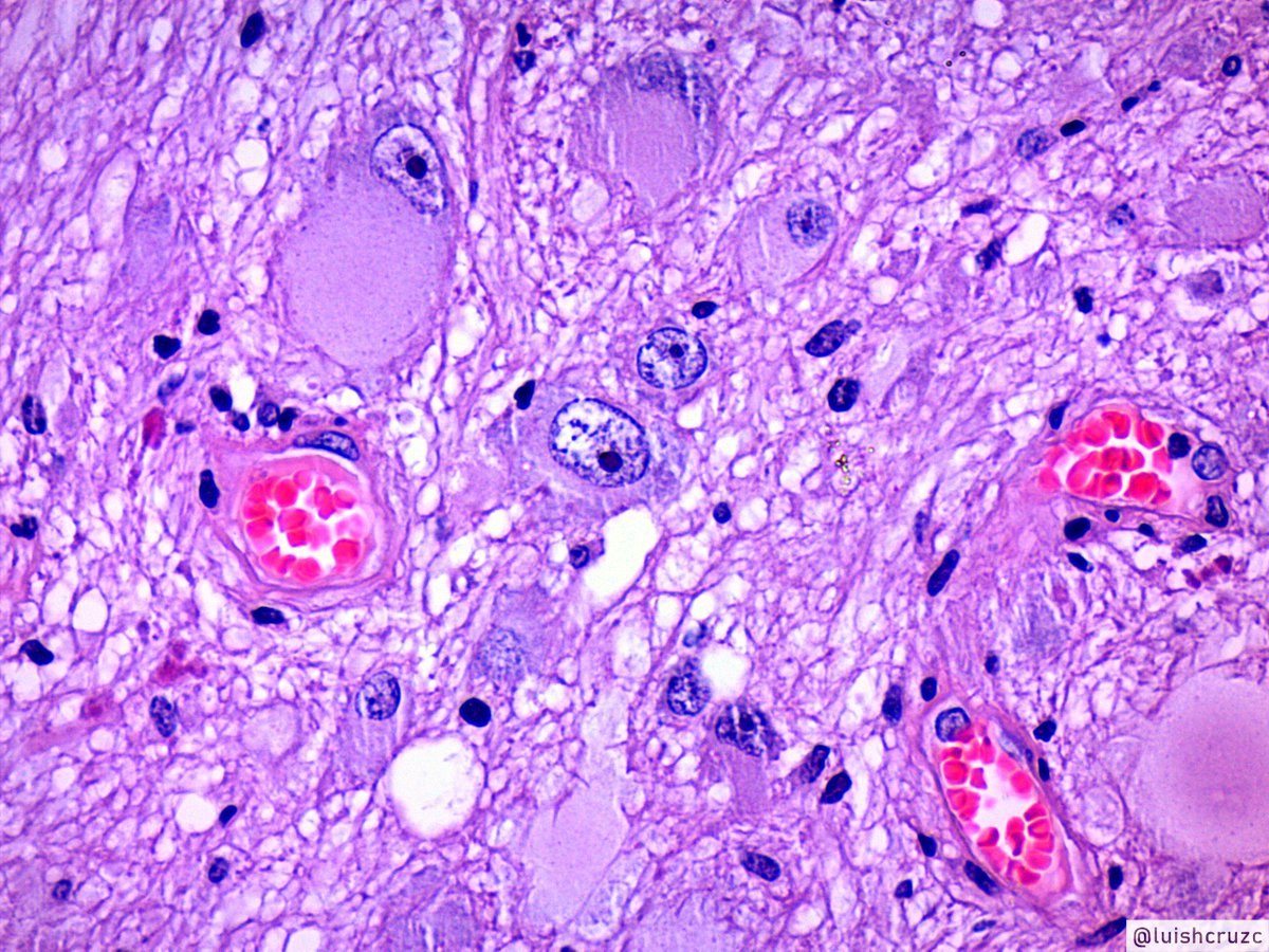
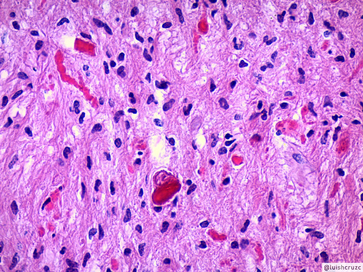
1/10 Ganglioglioma (GG)
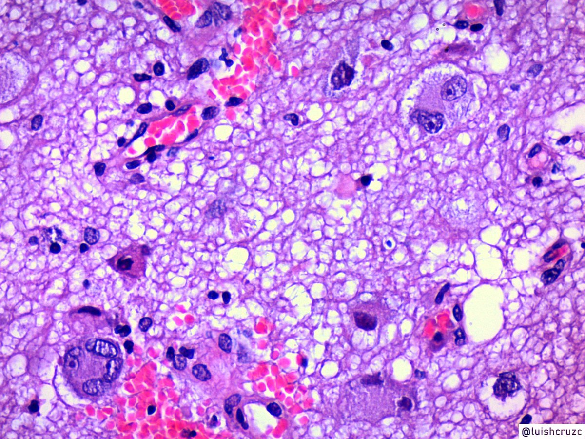
Mixed neuronal-glial tumor
🔵composed of dysplastic neurons and glial component
Others features are,
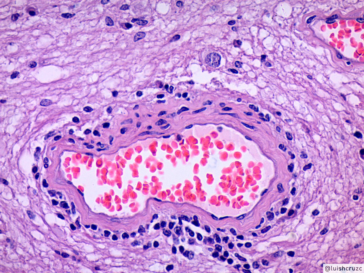
🔵Delicate vasculature
🔵lymphocyte cuffs around vessels
🔵Calcifications
#pathology #PediPath #neuropath #Tweetorial
Mixed neuronal-glial tumor
🔵composed of dysplastic neurons and glial component
Others features are,
🔵Delicate vasculature
🔵lymphocyte cuffs around vessels
🔵Calcifications
#pathology #PediPath #neuropath #Tweetorial
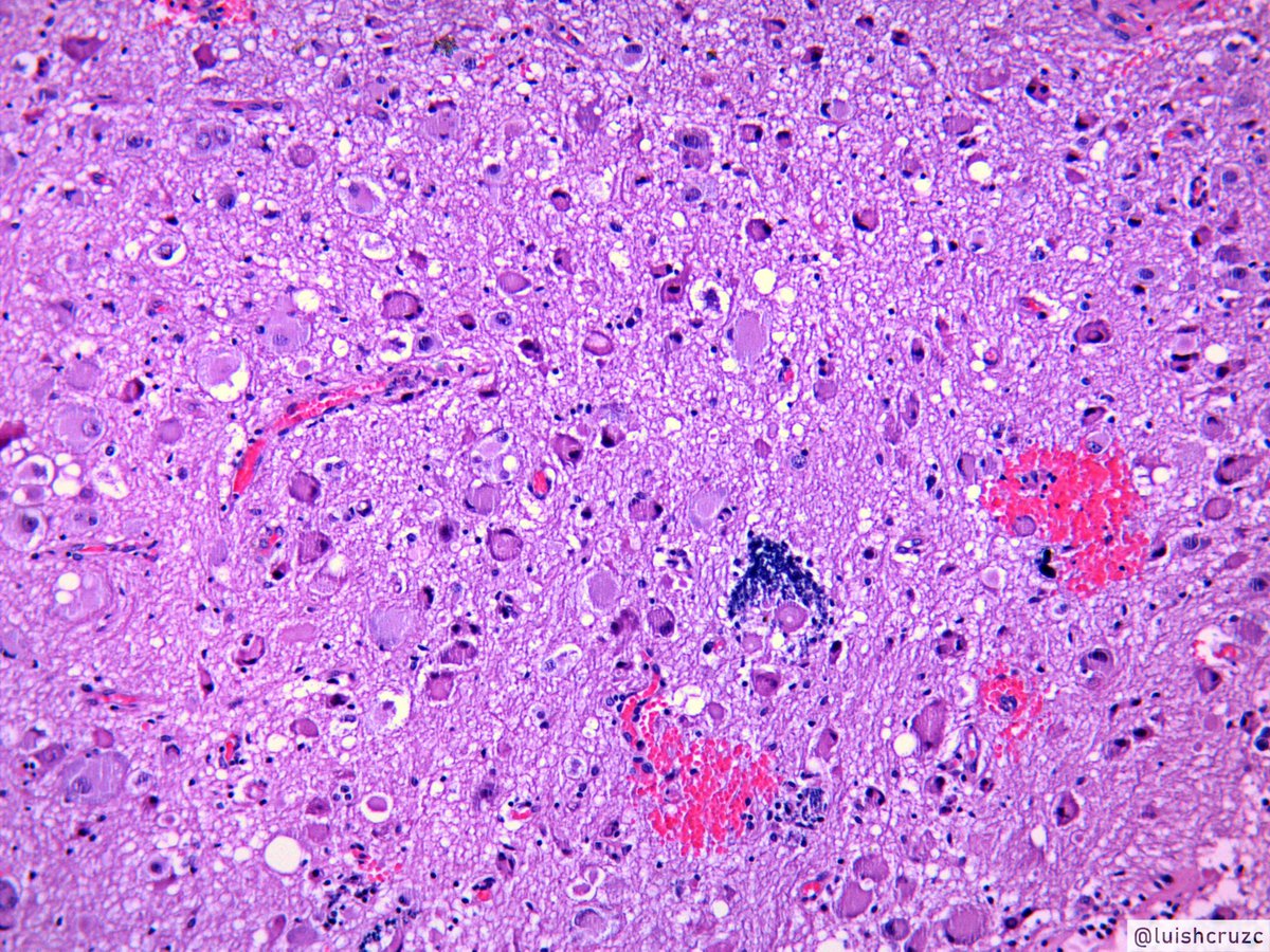

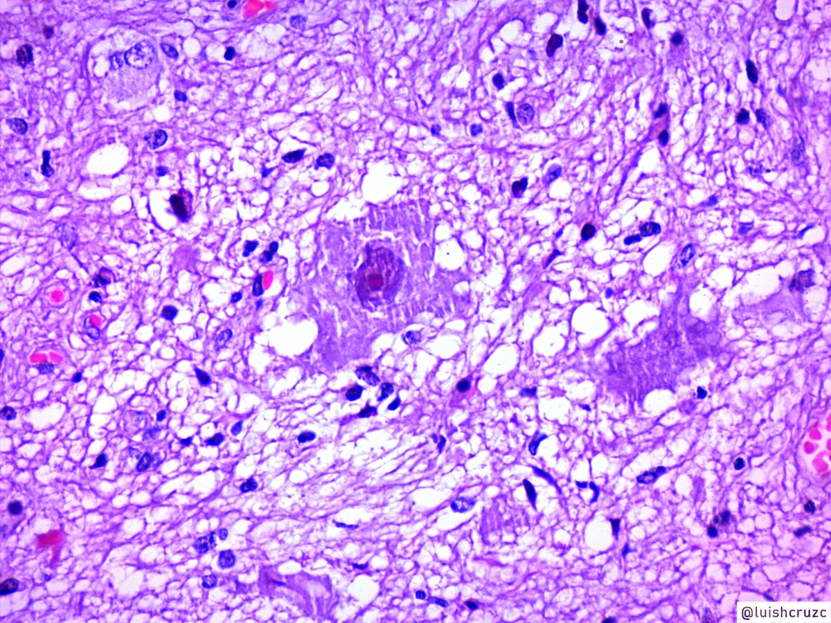
@sza_jhcyto @miguelguzmanmd @HopkinsNeuropth @edusqo @DrGeeONE @ariella8 @vi_monappa @BrainIsThePath @Path_Matt @JMGardnerMD 2/10 Glial component can resemble pilocytic astrocytoma with,
🔵Rosenthal fibers
🔵Eosinophilic granular bodies,
🔵oligodendroglioma and other gliomas-like components
#pathology #PediPath #neuropath
🔵Rosenthal fibers
🔵Eosinophilic granular bodies,
🔵oligodendroglioma and other gliomas-like components
#pathology #PediPath #neuropath


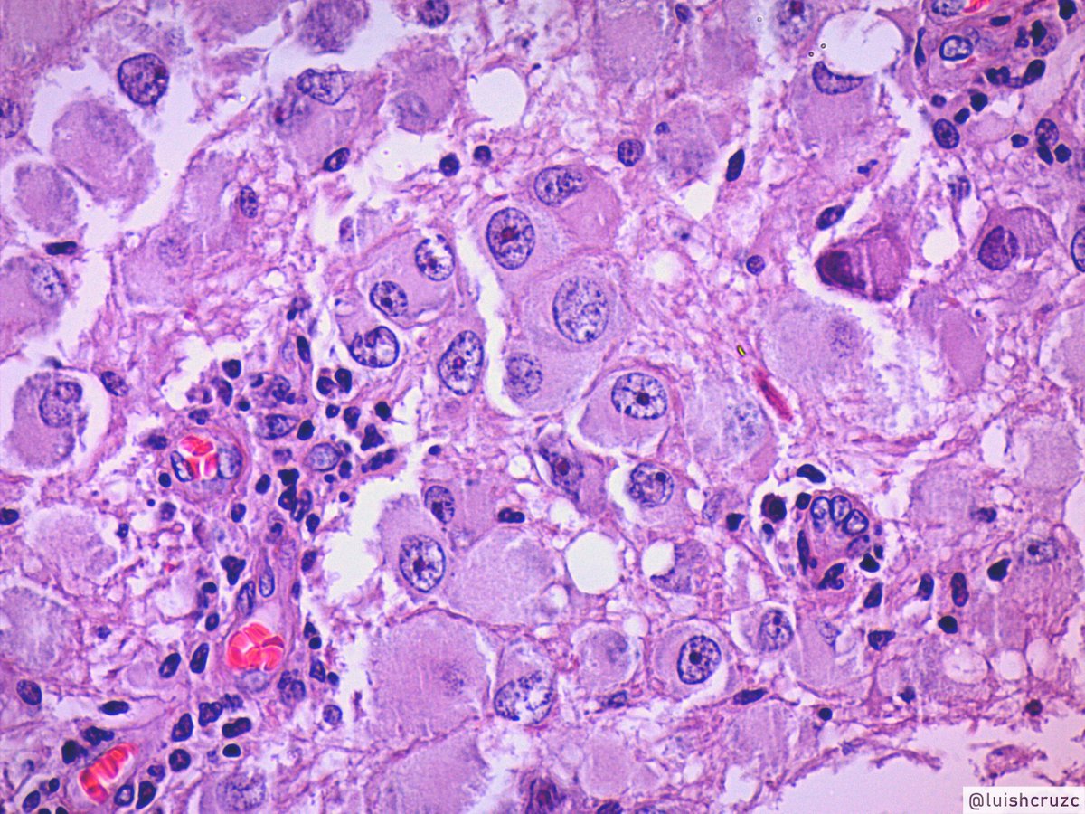
@sza_jhcyto @miguelguzmanmd @HopkinsNeuropth @edusqo @DrGeeONE @ariella8 @vi_monappa @BrainIsThePath @Path_Matt @JMGardnerMD 3/10 Neuronal component shows neurons with dysplasia
(dysmorphic neurons) characterized by:
🔵Clustering
🔵cytomegaly
🔵perimembranous aggregation of Nissl substance
🔵Binucleation
#pathology #PediPath #neuropath
(dysmorphic neurons) characterized by:
🔵Clustering
🔵cytomegaly
🔵perimembranous aggregation of Nissl substance
🔵Binucleation
#pathology #PediPath #neuropath
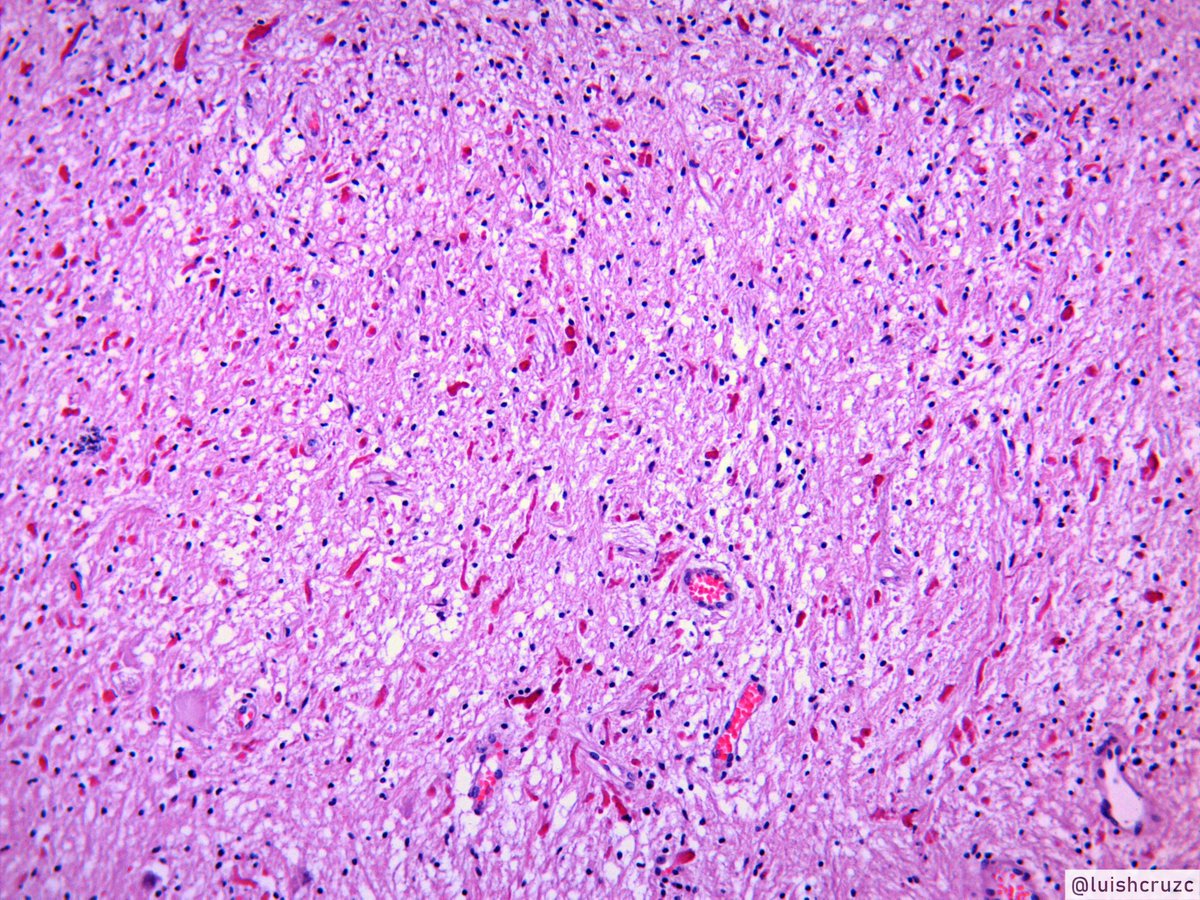
@sza_jhcyto @miguelguzmanmd @HopkinsNeuropth @edusqo @DrGeeONE @ariella8 @vi_monappa @BrainIsThePath @Path_Matt @JMGardnerMD 4/10 A note on Rosenthal fibers :
🔵eosinophilic cytoplasmic proteinaceous aggregates
🔵RFs are composed mainly of intermediate filaments & GFAP
🔵NOT specific for any tumor
🔵can also be found in reactive gliosis & neurodegenerative disorders(Alexander Disease)
🔵eosinophilic cytoplasmic proteinaceous aggregates
🔵RFs are composed mainly of intermediate filaments & GFAP
🔵NOT specific for any tumor
🔵can also be found in reactive gliosis & neurodegenerative disorders(Alexander Disease)
@sza_jhcyto @miguelguzmanmd @HopkinsNeuropth @edusqo @DrGeeONE @ariella8 @vi_monappa @BrainIsThePath @Path_Matt @JMGardnerMD 5/10
🔵Most GG are histologically WHO grade I, but some have anaplastic features in their glial component to be considered WHO grade III "anaplastic GG"
🔵enough mitosis, vascular endothelial proliferation or necrosis
🔵Strict criteria have not been established
#neuropath
🔵Most GG are histologically WHO grade I, but some have anaplastic features in their glial component to be considered WHO grade III "anaplastic GG"
🔵enough mitosis, vascular endothelial proliferation or necrosis
🔵Strict criteria have not been established
#neuropath
@sza_jhcyto @miguelguzmanmd @HopkinsNeuropth @edusqo @DrGeeONE @ariella8 @vi_monappa @BrainIsThePath @Path_Matt @JMGardnerMD 6/10
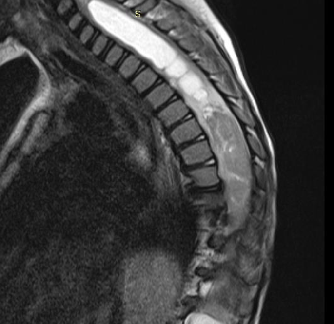
Imaging
usually well-demarcated, cystic with a mural nodule,
Can occur throughout the CNS
>70% occur in temporal lobe
here are some examples
Imaging
usually well-demarcated, cystic with a mural nodule,
Can occur throughout the CNS
>70% occur in temporal lobe
here are some examples
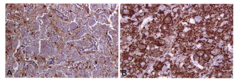
7/10 IHC will show positivity of neuronal markers such as synaptophysin, chromogranin, and neurofilament (in neurons)
and GFAP, .. in glial component
Not necessary for diagnosis
GFAP / Synaptophysin (photo from the WHO book)
#pathology #PediPath #neuropath
and GFAP, .. in glial component
Not necessary for diagnosis
GFAP / Synaptophysin (photo from the WHO book)
#pathology #PediPath #neuropath
8/10
CD34 can explore if it originated from cortical dysplasia, here are some references provided by @JCien_Path
CD34 can explore if it originated from cortical dysplasia, here are some references provided by @JCien_Path
@JCien_Path 9/10 Cortical dysplasia (Focal or diffuse)
Focal:
🔵region of the cerebral cortex that shows loss of laminar organization
🔵isolated large neurons
🔵 disorientation,
neurons, and widening of the cortex
Diffuse: Numerous regions
#pathology #PediPath
Focal:
🔵region of the cerebral cortex that shows loss of laminar organization
🔵isolated large neurons
🔵 disorientation,
neurons, and widening of the cortex
Diffuse: Numerous regions
#pathology #PediPath
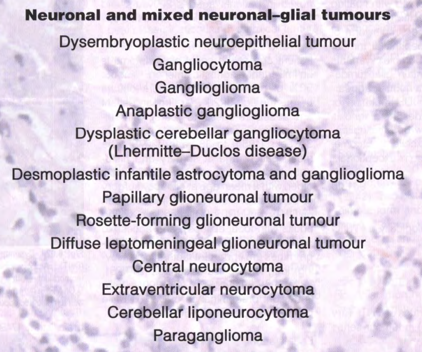
@JCien_Path 10/10 Other tumors in this category are:
(WHO Blue book)
End...
#pathology #PediPath #neuropath
(WHO Blue book)
End...
#pathology #PediPath #neuropath
@JCien_Path Inspired by the great tweetorial of @smlungpathguy . Harder than it looks, but worth it.