1/Welcome to our first OCT #Tweetorial! We'll cover a few key
concepts and share a step-by-step guide on the basics of image
interpretation 🤓 #imagefirst #OCTimaging
Important Safety Info: abbo.tt/2GyBrZ1
concepts and share a step-by-step guide on the basics of image
interpretation 🤓 #imagefirst #OCTimaging
Important Safety Info: abbo.tt/2GyBrZ1
2/First things first, what is OCT?
OCT stands for optical coherence tomography which is an imaging modality that uses near-infrared light to provide high definition images of the artery.
Important Safety Info: abbo.tt/2GyBrZ1
OCT stands for optical coherence tomography which is an imaging modality that uses near-infrared light to provide high definition images of the artery.
Important Safety Info: abbo.tt/2GyBrZ1
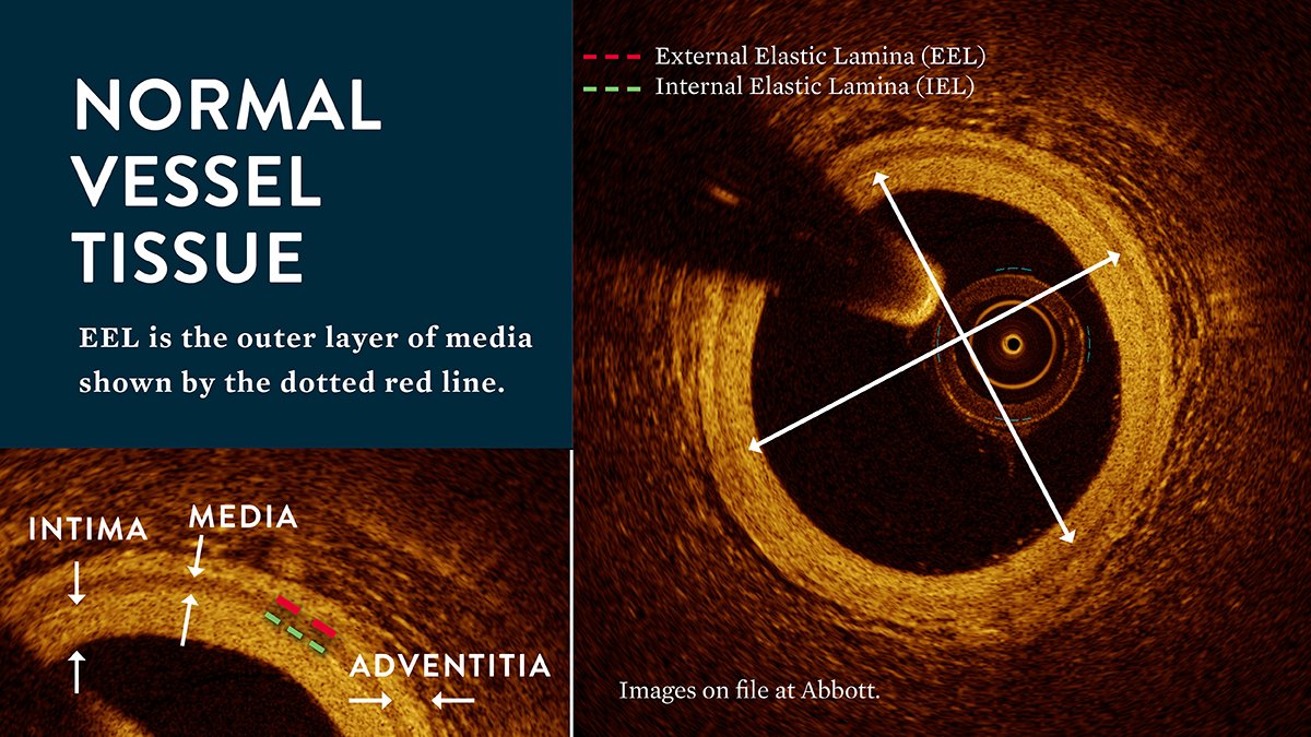
3/What are the basic elements of an OCT image? Here's what you see in a radial cross-sectional view:
Important Safety Info: abbo.tt/2GyBrZ1 #OCTImaging #imagefirst
Important Safety Info: abbo.tt/2GyBrZ1 #OCTImaging #imagefirst

4/Here's a view of normal vessel tissue. Media appears as a slightly darker band between the other two layers ("tire" looking dark line).
Important Safety Info: abbo.tt/2GyBrZ1#OCTIma… #imagefirst
Important Safety Info: abbo.tt/2GyBrZ1#OCTIma… #imagefirst
5/OCT helps to identify different microstructures, tissue and plaque
morphologies.
Here are the three most common types:
Important Safety Info: abbo.tt/2GyBrZ1 #OCTImaging #imagefirst
morphologies.
Here are the three most common types:
Important Safety Info: abbo.tt/2GyBrZ1 #OCTImaging #imagefirst
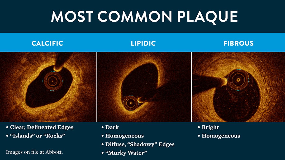
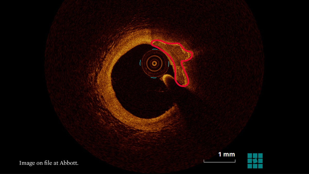
6/Here’s one way to remember visualization of most common plaque with OCT:
Fibrotic plaque = full of light
Lipid plaque = absence of light
Calcified plaque = a border can be drawn around it
Important Safety Info: abbo.tt/2GyBrZ1 #OCTImaging #imagefirst
Fibrotic plaque = full of light
Lipid plaque = absence of light
Calcified plaque = a border can be drawn around it
Important Safety Info: abbo.tt/2GyBrZ1 #OCTImaging #imagefirst
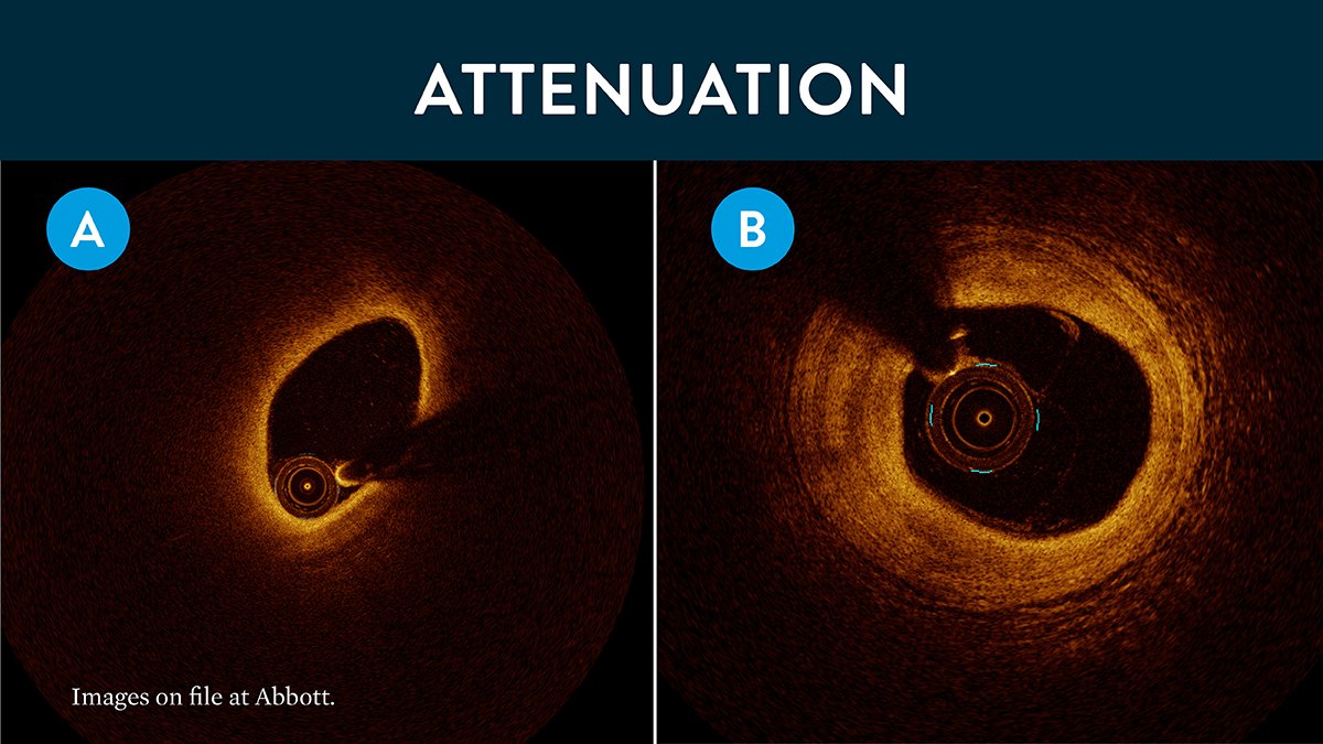
7/To describe images, we use "attenuation" in regards to light penetration, or
simply "loss of light"
(A) High attenuation=high loss of light, dark image, can't see deep tissue
(B) Low attenuation=light penetrates deep, can see tissue
Important Safety Info:abbo.tt/2GyBrZ
simply "loss of light"
(A) High attenuation=high loss of light, dark image, can't see deep tissue
(B) Low attenuation=light penetrates deep, can see tissue
Important Safety Info:abbo.tt/2GyBrZ
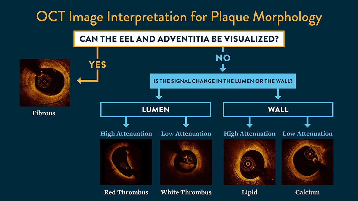
8/ To help you interpret OCT images, check out this cool algorithm developed by @ZiadAlinyc.
Simply put: can you see the EEL? And what do the lumen and vessel wall look
like?
Important Safety Info: abbo.tt/2GyBrZ1#OCTIma… #imagefirst
Simply put: can you see the EEL? And what do the lumen and vessel wall look
like?
Important Safety Info: abbo.tt/2GyBrZ1#OCTIma… #imagefirst