,
25 tweets,
10 min read
Read on Twitter
#Tweetorial time! 🚨 Looks like there’s another misconception about #cholesterol and #atherosclerosis making rounds again. This one has to do with the process of #LDL particles entering the arterial wall. So gather round friends and let me science the s*it out of this 🤓 (1/24)
For background, here’s a widely cited series of figures by Nakashima et al. (2007). They performed autopsies on 38 people aged 7-49, who died of non-#cardiovascular causes (2/24)
ncbi.nlm.nih.gov/pubmed/17303781
ncbi.nlm.nih.gov/pubmed/17303781
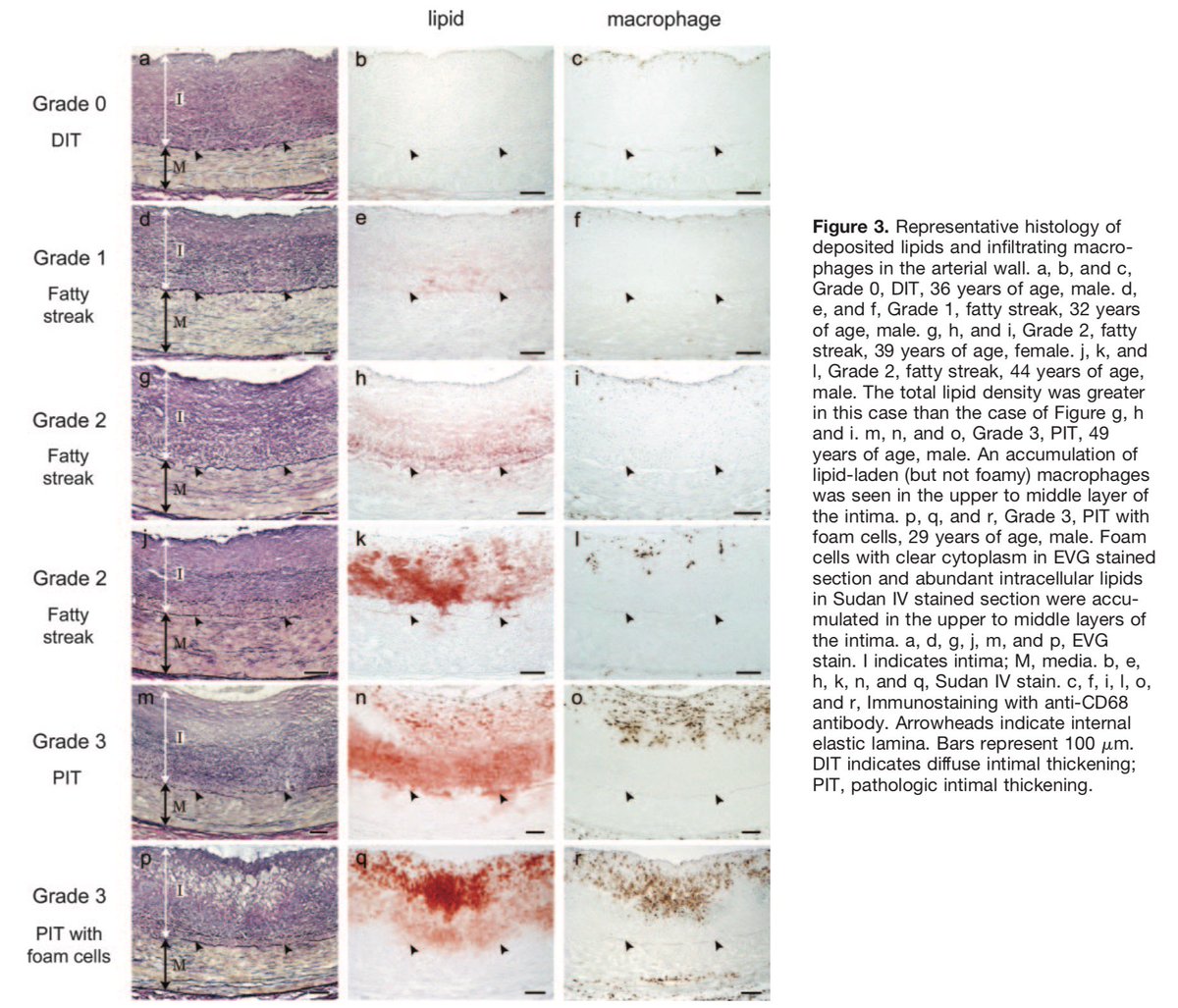
Nakashima et al. looked for atherosclerotic lesions at different stages, according to a previously published morphological classification scheme. They found them, took slices, stained them to find #lipids and #macrophages and organized them nicely (3/24)
(It should be noted that even though the famous figure series now looks like it could depict actual progression of a single plaque, individual figures are obviously from different subjects and different lesions at various stages) (4/24)
They present an elegant “time-series” of how lipids first appear in the vessel wall and then progress deeper and deeper eventually pooling into one great big mass. Lipids are followed by macrophages - cells that eat them (5/24)
This is a GREAT illustration of the current paradigm of atherogenesis (“response to retention”), meaning that LDL particles enter the arterial wall, get trapped, aggregate & become inflammatory. NOT some other sequence of events: ncbi.nlm.nih.gov/pubmed/7749869 (6/24)
So here’s the topic of this tweetorial: according to some, there’s something fishy in this series! Let’s look at the first image. Notice how the lipid stains appear quite far from the inner layer of the wall (endothelium)? OMG HOW DO THEY “JUMP” THERE!? (7/24)

Now, if you’re the arrogant type at the peak of #DunningKruger Effect (AKA “Mount Stupid”), you might think “Gotcha!! I read this and now I know better than all the experts in the world! Clearly this disproves 100 years of CVD research!”. And you’d be wrong (8/24)
On the other hand, if you’re a more moderate, critical thinker but a curious person, you might go “Huh, that’s interesting but I’m probably I’m missing something. I’m sure these folks who do this for a living have thought about this”. And then you’d be right! 😊 (9/24)
So what is the explanation? Before we get there, let’s take a quick poll: without checking, what do you think is the *scale* of the Nakashima images? (10/24)
Now, what do you think is the size scale of #LDL lipoproteins?
(11/24)
(11/24)
Now that we’re done polling, let’s get back to the paper and take a look at the scale bar in the corner of each fig. It’s 100 MICRO meters. Then, let’s remember that LDL particles are in the 20-30 NANO meter range. There’s a THOUSAND fold difference in scale! (12/24)
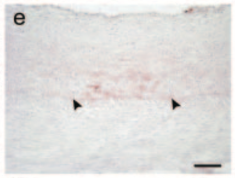

So the short answer to the “jump mystery" is simply image resolution. There ARE indeed #LDL particles in the area just below endothelium but they’re just too small to be seen. If Nakashima et al. had used a microscope with 800x magnification, they might have caught those! (13/24)
Think of it this way: essentially the situation is analogous to looking at a satellite image of a city at a scale of individual blocks. At best, the resolution allows you to see large aggregate masses of people but not individual humans (14/24)

Speaking of mass, back to red stuff. The authors are using a stain called Sudan IV, which turns neutral lipids (triglycerides etc) red. The accumulating redness is the build-up and fusion of nm-sized LDL particles to such a degree it starts to show up in micrometer-scale (15/24)

“But why does it build up like that?” Great question, glad you asked! Aside from the scale bars, we now have to note the little arrows. They indicate something called Internal Elastic Lamina (IEL). It’s right at the border between two layers of the wall (intima and media) (16/24)


For many macromolecules, IEL is an impermeable barrier. It has holes which are small enough for some HDL particles or albumin to squeeze through but LDLs simply don’t fit. This was shown in seminal work already in the 80’s: ncbi.nlm.nih.gov/pubmed/6161619 (17/24)
(BTW, that paper is related to a line of research showing how the concentration of LDL particles in the wall is more than two-fold to their concentration in blood. The wall is a sticky environment to them and that’s why we don’t want them getting there in the first place) (18/24)
So the red mass shown by Nakashima is essentially a massive amount of LDL particles hitting a wall and forming one great big pile of lipid that stains red! Any questions? Yes, you in the back (19/24)
“But how can we be sure that there REALLY are individual LDLs in the layer just under the endothelium? Are there any studies that HAVE used a sufficiently big magnification that would reveal them?” Another great question! Answer is: yes (20/24)
@Salehti et al. have used a 3D electron microscope to study small LDL deposits and cholesterol crystals in human carotid arteries. It’s a very nice paper with heaps of interesting data! ncbi.nlm.nih.gov/pubmed/29154769 (21/24)
Among others, the paper contains this supplemental figure that clearly shows actual cholesterol CRYSTALS just over 100um from the endothelium (in the Nakashima paper, the red starts to build up around 300-400um) (22/24)

So, in conclusion, the people who look at the Nakashima figure and think there’s something dubious about it that scientists haven’t noticed, are missing the scale difference of plaques and lipoproteins. Scientists investigating atherosclerosis are not stupid (23/24)
ApoB-containing lipoproteins are travelling in the bloodstream and end up into the arterial walls from there. The more particles, the higher the risk of a build-up over time. That’s what’s shown again and again in multiple lines of evidence that you should believe (24/24)
Big thanks to @Salehti for clarifying a few things related to this! Please give her a follow if you’re interested in following an actual expert in lipoprotein infiltration to the arterial wall.
/END tweetorial (25/Bonus)
/END tweetorial (25/Bonus)