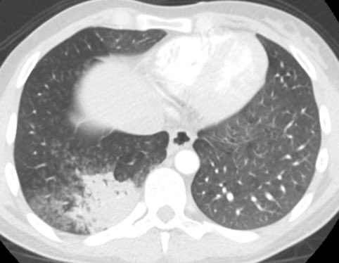
If report CT chest, this is an important entity to know about. This is a very good example of it. Not quite middle-aged male with chest pain and fever, no other medical history.
What is your diagnosis?
#FOAMrad #Radres
@JeffreyKanneMD @CsFuss
What is your diagnosis?
#FOAMrad #Radres
@JeffreyKanneMD @CsFuss
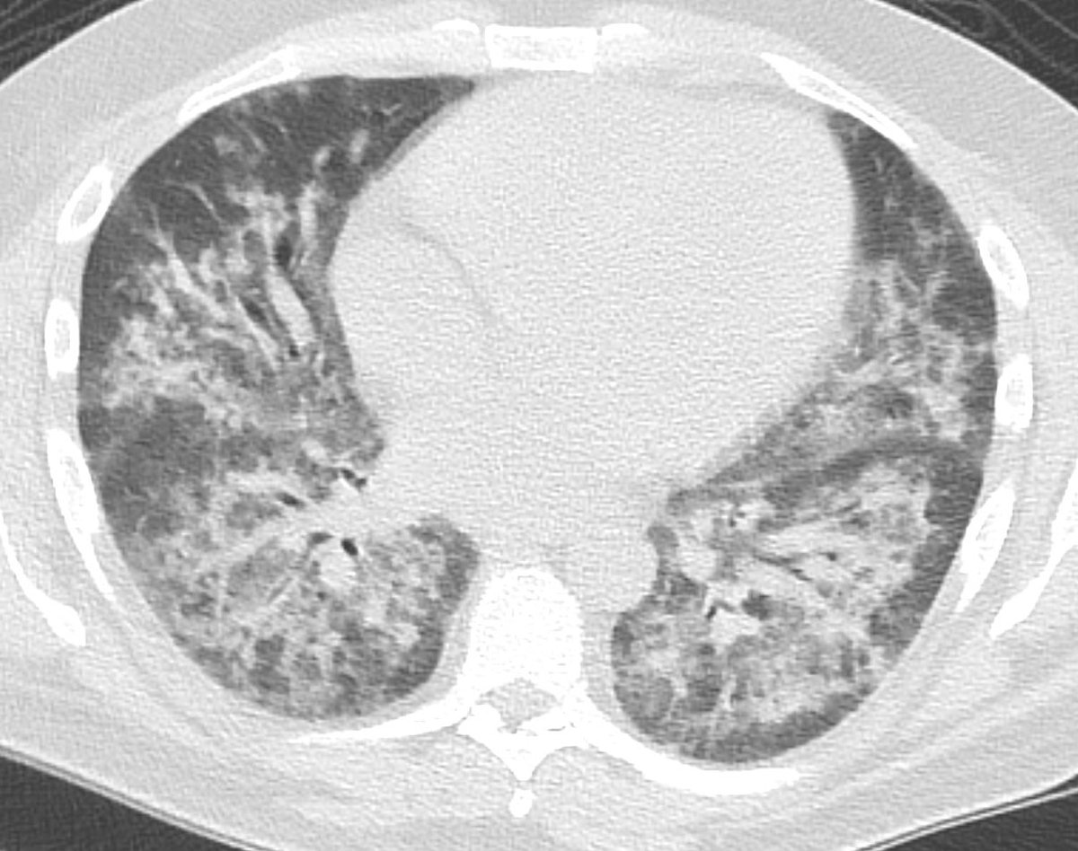
Here are the lung windows, showing pretty typical right lower lobe pneumonia. Does this change your thinking about the first image? Note that there is no interstitial edema in the RLL or pleural effusion.
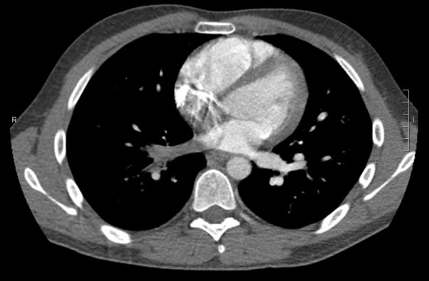
This is the coronal MIP image. Notice how there is decreased pulmonary vascularity in the right lung vs left lung. Remembering pulmonary physiology? There is increased pulmonary arterial resistance in areas with low alveolar oxygen levels, to shunt blood away from low V/Q.

So looking again at our first image, the absence of contrast in the right inferior pulmonary vein is simply unenhanced blood rather than thrombus. There is a delay in contrast enhancement due to slower flow through the right lung as a physiologic response to pneumonia.

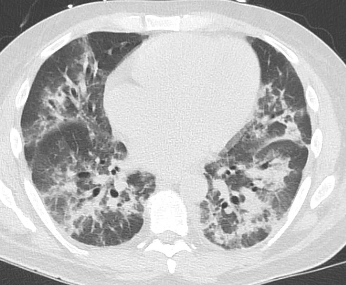
If we look at lung windows, decreased perfusion through the right lung can be appreciated as diffusely decreased density of the right lung. Interestingly this involves much more lung than just the area of pneumonia - here much of the right lung is affected.

Might there still be thrombus in the pulmonary vein? It’s very unlikely. Pulmonary vein embolism occurs but is very rare, and usually happens after lung transplant, cardiac surgery, or pneumonectomy, or after RF ablation for a fib.
This patient did not have any of these.
This patient did not have any of these.
Another important thing to think about here - if this was a big thrombus in the right inferior pulmonary vein, there would be significant interstitial edema/septal thickening in the right lower lobe, and probably an effusion. These are not present.
In this case I would have comfortably called this artifact. If you aren’t sure, you can get another CT in the venous phase, which I would recommend prior to procedure/hospital transfer if you are fixing to diagnose pulmonary vein thrombus in a low risk patient!
Here is a link to a terrific review of pulmonary vein imaging - a great reference to have on hand:
Unfortunately could not find authors to tag them on twitter :-).
End.
doi.org/10.1148/rg.201…
Unfortunately could not find authors to tag them on twitter :-).
End.
doi.org/10.1148/rg.201…