,
26 tweets,
9 min read
Read on Twitter
*thread alert*
This is your host (@MySci_PK) and I will tell you all about our latest publication which I'm super duper proud of!
This is your host (@MySci_PK) and I will tell you all about our latest publication which I'm super duper proud of!
*drumroll*
This publication is a result of most of my PhD work.
This publication is a result of most of my PhD work.

Let us start with breaking down the keywords
1. transparent
2.graphene
3. PEDOT:PSS
4. microelectrodes for electrophysiology
1. transparent
2.graphene
3. PEDOT:PSS
4. microelectrodes for electrophysiology
1. transparent
We mean optically transparent, just like window glass (or even better 😉)
Don't get carried away by the screen in the gif. Stay with me!
We mean optically transparent, just like window glass (or even better 😉)
Don't get carried away by the screen in the gif. Stay with me!
2. graphene
I talked a lot about this wonder material of the 21st century. In case you missed it, here is the thread 😁
I talked a lot about this wonder material of the 21st century. In case you missed it, here is the thread 😁
3. PEDOT:PSS
poly(3,4‐ethylenedioxythiophene) polystyrene sulfonate
is a conductive polymer used in the microelectrode array technology to improve the electrical property of the microelectrodes.
poly(3,4‐ethylenedioxythiophene) polystyrene sulfonate
is a conductive polymer used in the microelectrode array technology to improve the electrical property of the microelectrodes.

4. Microelectrodes for electrophysiology
The details about microelectrode arrays are here:
The details about microelectrode arrays are here:
After familiarising the keywords in the title, let us dig deeper and go through the published work.
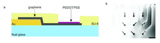
#graphene was produced following chemical vapour deposition and integrated in the MEA technology (details in the thread)
(a) Side view of the MEA (not to scale)
(b) top view scanning electron microscopic image of one of the electrode fields. Bright - Au conduction paths covered with graphene
dark - graphene
(b) top view scanning electron microscopic image of one of the electrode fields. Bright - Au conduction paths covered with graphene
dark - graphene
PEDOT:PSS (the conductive polymer) was the magic which the graphene microelectrodes needed to improve the electrical properties.
We didn't really sprinkle PEDOT:PSS on graphene as seen in the gif. What we did is in the next tweet!
We didn't really sprinkle PEDOT:PSS on graphene as seen in the gif. What we did is in the next tweet!
PEDOT:PSS (magic dust) was electrodeposited on the transparent graphene microelectrodes.
(a) Three-electrode set up during the electrodeposition
(b) The amount of PEDOT:PSS on the graphene was controlled by the length of the process.
Longer = more PEDOT:PSS
(a) Three-electrode set up during the electrodeposition
(b) The amount of PEDOT:PSS on the graphene was controlled by the length of the process.
Longer = more PEDOT:PSS

Now, I know what you are thinking. Graphene is transparent, but what happens when PEDOT:PSS comes into the picture?
Won't the transparency go for a toss?
We thought the same!
Won't the transparency go for a toss?
We thought the same!
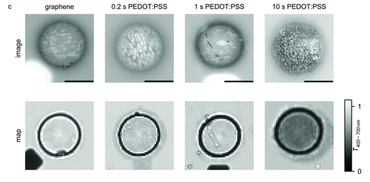
Here are the optical transmittance maps for 4 different microelectrodes.
Just to give you an idea, the optical maps (2nd row) took 12-15 hr each.
Just to give you an idea, the optical maps (2nd row) took 12-15 hr each.

Also, every pixel in the optical map is ~1 µm. That is 0.000 000 001 km.
that is how small and precise the measurement was.
The goal was correlate the optical map with the optical image.
And boy, was it beautifully achieved? I would say so! 😊
that is how small and precise the measurement was.
The goal was correlate the optical map with the optical image.
And boy, was it beautifully achieved? I would say so! 😊
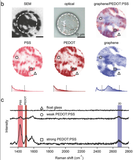
Now onto the result I am extremely proud of - Raman Spectroscopy
Here are Raman spectra of three different electrodes.
Here are Raman spectra of three different electrodes.

The PEDOT, PSS and graphene (2D) peaks are highlighted.
The areas under the peaks in the highlighted range was integrated and plotted which produced Raman map (next tweet)
The areas under the peaks in the highlighted range was integrated and plotted which produced Raman map (next tweet)

Raman maps correlated with the scanning electron and optical microscopy
Let us go through the images one by one.
SEM: dark - graphene, bright - substrate (glass)
optical: after PEDOT:PSS electropdeposition, brown - PEDOT:PSS
Let us go through the images one by one.
SEM: dark - graphene, bright - substrate (glass)
optical: after PEDOT:PSS electropdeposition, brown - PEDOT:PSS

graphene/PEDOT:PSS map is a superimposition of individual graphene, PEDOT, PSS maps seen in the 2nd row
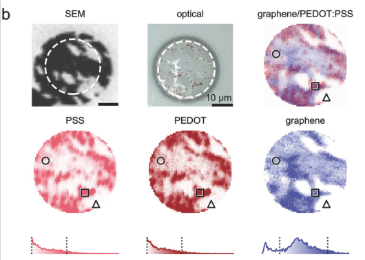
How do the Raman spectra look for different regions? 👇
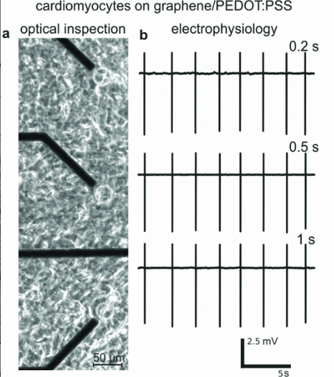
(a) happy healthy beating heart cells cultured in the lab. The heart cells on top of the microelectrodes are visible.
optical investigation ✅
optical investigation ✅

(b) The health and happiness of the heart cells is confirmed by electrophysiological recordings.
Every vertical line in (b) is a heart-beat. 😁
Every vertical line in (b) is a heart-beat. 😁
I probably should have lead with this. 🤪
this is the WHY to the need for transparent microelectrodes in the MEA technology.
this is the WHY to the need for transparent microelectrodes in the MEA technology.
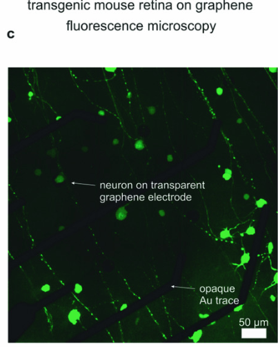
And now to my favourite favourite image! This picture sums up my PhD work story really well.
Visualising neurons through the transparent graphene microelectrodes, while the Au conduction paths (black lines) block the view.
#Win
Visualising neurons through the transparent graphene microelectrodes, while the Au conduction paths (black lines) block the view.
#Win
Thank you for staying till the end!
In case of questions, do reach out on @MySci_PK and I will be more than happy to have a conversation about graphene, MEAs, transparent microelectrodes, electrophysiology and other topics. 😊
*The End*
In case of questions, do reach out on @MySci_PK and I will be more than happy to have a conversation about graphene, MEAs, transparent microelectrodes, electrophysiology and other topics. 😊
*The End*