
,
85 tweets,
21 min read
Read on Twitter
In this thread (continued), I am looking back at highly cited (over 1000 citations) 'historical' (prior to 2008) bionano papers, in particular those that deal with nanoparticles & cells. Comments most welcome. #bionano #NanoBubble.
Some background on selection of papers + comments on the first 10 papers here
threadreaderapp.com/thread/1093179…
threadreaderapp.com/thread/1093179…
#11 2005, El-Sayed et al "Surface Plasmon Resonance Scattering and Absorption of anti-EGFR Antibody Conjugated Gold Nanoparticles in Cancer Diagnostics: Applications in Oral Cancer"
doi.org/10.1021/nl0500…
doi.org/10.1021/nl0500…
That paper uses two types of gold nanoparticles: 1) conjugated w/ EGFR Ab, or, 2) not conjugated with an antibody.
And three epithelial cell lines : one non-cancerous (HaCat) and two cancerous (HOC and HSC).
And three epithelial cell lines : one non-cancerous (HaCat) and two cancerous (HOC and HSC).
First comment: no measurement comparing EGFR expression in these 3 cell lines. They seem to assume that "non-cancerous" means no EGFR. As a matter of (known at the time) fact, HaCat do express EGFR:
ncbi.nlm.nih.gov/pubmed/10527633
ncbi.nlm.nih.gov/pubmed/10527633
The paper has three figures. All of them are dark field microscopy images, with some additional spectroscopy panels added for figures 2 and 3.
Fig 1 is dark field microscopy (or "light scattering image") of unlabelled cells. It is a useful reference for the next two figures; but still kind of fascinating that a simple microscopy image of cells makes a full article figure given that...
dark field microscopy made its first appearance in the scientific literature in 1848, and had been used to observe biological samples well before the publication of "Modern Dark-Field Microscopy and the History of Its Development"... in 1920.
biodiversitylibrary.org/page/27095464#…
biodiversitylibrary.org/page/27095464#…


Back to Fig 1: ".. we can see tht the 3 types of cells hve ≠ structure characteristics. HOC cancer cells r almost 4 times larger than HaCaT or HSC cells. HaCaT & HSC cells show almost homogeneous diamond shapes while HOC cells have other shapes for some cells." 🤨🤔

👆 Can you see the diamonds? 💎💎💎
More seriously, what strikes me in this short description is how vague and unscientific the language is. It also suggests that the authors are not very familiar with looking at cells in a microscope.
More seriously, what strikes me in this short description is how vague and unscientific the language is. It also suggests that the authors are not very familiar with looking at cells in a microscope.
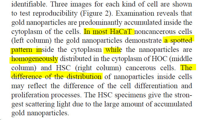
Fig 2 is similar experiment but where the cells have been exposed to unconjugated gold nanoparticles (35 nm diameter, 0.3 nM, 48h). The figure legend says that it shows that there is no difference btw the cells, but several paragraphs of text discuss supposed differences. 🤔


Finally, comes Fig 3... Quite simply, can you see the cells? If you do, are any
💎-shaped?
Seriously, am I the only one finding this crazy? I have no idea what the first column shows, and my best guess is that the next two are dead cells.
💎-shaped?
Seriously, am I the only one finding this crazy? I have no idea what the first column shows, and my best guess is that the next two are dead cells.

This paper 👆 has been cited 1942 times according to Google Scholar.
I thought I had finished with El-Sayed et al, #11, but there is more 😲🥴.
The press release, and the Georgia tech abstract, say that ".. researchers found that the gold nanoparticles have 600 percent greater affinity for cancer cells than for noncancerous cells."
The press release, and the Georgia tech abstract, say that ".. researchers found that the gold nanoparticles have 600 percent greater affinity for cancer cells than for noncancerous cells."
Where does this 600% comes from?
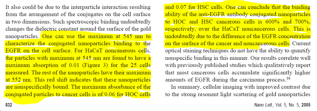
The "600% greater affinity than to the noncancerous cells" comes from spectroscopy in fig 3.
Let's ignore that I can't actually see cells in Fig 3. (see 👆).
Let's also ignore that there is no affinity measurement in this paper (that would require a titration).
Let's ignore that I can't actually see cells in Fig 3. (see 👆).
Let's also ignore that there is no affinity measurement in this paper (that would require a titration).
Here is the spectroscopy in Fig 3. Each spectrum is supposed to represent a different cell. Now, if you give it a quick look, you might conclude that there is roughly the same amount of material in each lines, with cell-to-cell variability in each case?

The authors however see something very different because they compare THIS ONE with THOSE ONES. And yes, there is a factor 6 between those, QED.

This is their justification. Totally ridiculous.
#12 is coming... sorry for the interruption.
Hussain et al, 2005, In vitro toxicity of nanoparticles in BRL 3A rat liver cells.
"This study was undertaken to address the current deficient knowledge of cellular response to nanosized particle exposure."
Hussain et al, 2005, In vitro toxicity of nanoparticles in BRL 3A rat liver cells.
"This study was undertaken to address the current deficient knowledge of cellular response to nanosized particle exposure."
6 figures
≠ NP sizes
≠ materials silver (Ag; 15, 100 nm)
molybdenum (MoO3; 30, 150 nm)
aluminum (Al; 30, 103 nm)
iron oxide (Fe3O4; 30, 47 nm)
titanium dioxide (TiO2; 40 nm)
≠ NP sizes
≠ materials silver (Ag; 15, 100 nm)
molybdenum (MoO3; 30, 150 nm)
aluminum (Al; 30, 103 nm)
iron oxide (Fe3O4; 30, 47 nm)
titanium dioxide (TiO2; 40 nm)
Also "relatively larger particles of" [ie not nanoparticles]:
cadmium oxide (CdO; 1 μm)
manganese oxide (MnO2; 1–2 μm)
tungsten (W; 27 μm)
cadmium oxide (CdO; 1 μm)
manganese oxide (MnO2; 1–2 μm)
tungsten (W; 27 μm)
I know I should not get distracted, but I can't help tracking the origins of this myth according to which NPs magically cross the cell membrane, so, from the intro:
"The major tox concern is [...] and some particles transport across cell membranes esp into mitochondria (Foley et al, 2002 dx.doi.org/10.1016/S0006-…)."
Foley et al is a single figure paper (about C60 which for a time was supposed to revolutionise drug delivery). Convinced?
Foley et al is a single figure paper (about C60 which for a time was supposed to revolutionise drug delivery). Convinced?

Back to #12, Hussain et al. Fig 1 and Fig 2 establish, using two different tox assays (LDH leakage and MTT) that at doses up ~ 100 µg/mL of particles, there is no toxicity in rat liver cells (BRL 3A) except for Ag (and for CdO which they use as "positive control").
The rest of the article (Figs 3-6) focuses on silver nanoparticles.
Here is what Fig 3 is supposed to show:

And here is the evidence, Fig 3 itself.
Can you see the "morphology of the cells"? Can you distinguish those particles that are associated with "cells" versus those associated with "membranes"?
Well, I certainly can't.
Can you see the "morphology of the cells"? Can you distinguish those particles that are associated with "cells" versus those associated with "membranes"?
Well, I certainly can't.


Fig 4 shows an increase of reactive oxygen species as a function of Ag nanoparticles concentration (using DCFH-DA, ie a fluorescent probe).
Fig 5 shows a decrease of mitochondrial membrane potential as a function of Ag nanoparticles concentration (using rhodamine 123, ie a fluorescent probe).
Fig 6 shows GSH depletion. The assay "Glutathione Assay Kit (Cayman Chemical Company, Ann Arbor, MI" relies on measuring absorbance at 405 or 412 nm which is potentially problematic as this is also the max absorbance of Ag NPs...
re last tweet, although correct, thinking about it, this would probably reduce the effect seen - hig abs corresponds to high GSH (caymanchem.com/product/703002) whereas they conclude [and that seems plausible] that Ag NPs deplete GSH.
So, apart from Fig 3, I do not have major criticism of this article, but hey, look at the conclusion:
"In summary nanoparticles lead to cellular morphological modifications"
What? Where did #12 show this? Only evidence on morphological modif is for Ag NPs (fig 3) & is v poor. In any way, Ag NPs are toxic; when cell dies they do change morphology. That's due to cells dying.
What? Where did #12 show this? Only evidence on morphological modif is for Ag NPs (fig 3) & is v poor. In any way, Ag NPs are toxic; when cell dies they do change morphology. That's due to cells dying.
The entire conclusion is written as a general truth about "nanoparticles" when it should be specifically and only be about silver nanoparticles, which according to the authors' own data behave pretty differently from the other materials studied.
Worse, the authors' own data suggest that the results have very little to do about nanoparticles and instead about the amount of Ag: the 15 nm and 100 nm particles give exactly the same results (Fig 4-6 below).


#13, Kirchner et al, Cytotoxicity of Colloidal CdSe and CdSe/ZnS Nanoparticles dx.doi.org/10.1021/nl0479…
"Cytotox of CdSe & CdSe/ZnS nanoparticles has been investigated 4 ≠ surface modifs such as coating w/ mercaptopropionic acid, silanization, & polymer coating"
"Cytotox of CdSe & CdSe/ZnS nanoparticles has been investigated 4 ≠ surface modifs such as coating w/ mercaptopropionic acid, silanization, & polymer coating"
Three figures.
Fig 1 shows that whatever the coating and size, particles enter cells and "ingested particles are stored in vesicular structures around the nucleus". Yep.
Fig 1 shows that whatever the coating and size, particles enter cells and "ingested particles are stored in vesicular structures around the nucleus". Yep.

Fig 2 shows the ratio R between the number of adherent cells after vs before incubation with NPs or Cd salts.
They do the nanoparticles incubation (18 h) in serum free "SATO medium"... which in and of itself blocks cell division + cause some cells to detach : R always ‹ 1
They do the nanoparticles incubation (18 h) in serum free "SATO medium"... which in and of itself blocks cell division + cause some cells to detach : R always ‹ 1

It is a bit worrying that at the lowest concentrations tested, one does not get the same R value for all conditions since it should correspond to no effect.
The trough shape of the curve -_- is Non-toxic (R constant) then Toxic (R decreases) then So-Toxic-Cells-Don't-Have-Time-To-Detach
Overall the paper makes a convincing case that the toxicity depends on the dissolution and that the coatings [i.e. the ones used in this study] play a role probably both in terms of affecting dose, via uptake, and possibly directly.
The latter is based on the observation of toxicity with coated gold particles (which is surprising and can't be due to ions).
Fig 3 is a "negative result" experiment where it is claimed that, at low dose (10 nM MPA-coated CdSe/ZnS particles) do not affect "characteristic electrophysiological properties of the cells."
This conclusion arises from patch-clamp recordings "under identical experimental circumstances of untreated and incubated cells" - they use two cell lines.
I don't really understand the point of this experiment (for which there is no NP conc dependence measured). Also, no statistics.
#14 Loo et al, Immunotargeted Nanoshells for Integrated Cancer Imaging and Therapy
dx.doi.org/10.1021/nl0501…
dx.doi.org/10.1021/nl0501…
"Here we provide an in vitro demonstration of the dual imaging/therapy approach, first detecting and then thermally ablating human breast cancer cells that overexpress HER2 using immunotargeted nanoshells that have been designed to both scatter and absorb light within the NIR"
The paper has three figures.
Fig 1 is abt materials prep as u wld expect: "Shell thickness was mathematically corroborated by Mie scattering theory w/ good agreement w/ SEM. Fig 1 shows the spectral characteristics & SEM image of nanoshells possessing a 10 nm thick shell that were used in this study"... But
well, look carefully at Fig 1 & its legend: the experimental spectra are not shown, just the theory. And the SEM is one image of one particle, at a resolution which makes it completely impossible to evaluate the shell thickness.

There is no supporting information. As far as I can see, this one SEM image of one nanoshell is the full extent of materials characterisation in this paper.

Fig 2-3 shows that cells exposed to the antibody-linked nanoshell + irradiation leads to cell death whereas non-specific antibody, or no irradiation, results in no toxicity. It is OK... but so little data! No characterization of the amount of uptake nor of the abs of cells.
Loo et al has been cited 1906 times according to Google Scholar.
#15 Morones et al, The bactericidal effect of silver nanoparticles
doi.org/10.1088/0957-4…
(#12 and #9 were also about silver NPs)
doi.org/10.1088/0957-4…
(#12 and #9 were also about silver NPs)
"Silver ions have been demonstrated to be useful and effective in bactericidal applications, but due to the unique properties of nanoparticles nanotechnology presents a reasonable alternative for development of new bactericides."
As noted in my discussion of #9, this is not new.
But let's carry on.
But let's carry on.
Presumably, given the title, you might think that this paper is about silver nanoparticles interacting with bacteria. I guess most of the 1,000 of articles citing it think so too. I am not so sure.

There is a characterization figure with a bizarre discussion of how some nanoparticles are extracted from this carbon matrix by the electron beam. But no characterization of proportion or time-dependence of NPs leaving the matrix is shown.
So, for all we know, it could be the "powder of silver nanoparticles inside a carbon matrix" which interact w/ the bacteria, then releasing silver ions. According to the inset of Fig 1a, these powder particles are in the micrometer range.

OK, at that point, I'd like to call proper electron microscopists to have a look at this paper. I am generally puzzled but I would really appreciate if any microscopist could comment regarding the extent to which the images and analysis support the conclusions. #realtimechem
Though I am still going to make a comment about the main conclusion, also reproduced in the abstract, which, I suspect, is the main cause of the astronomic number of citations.

Nano is cool because size dependence gives special property. This is the key winning message. It's right there in this abstract. That is of course quite an easy thing to test. Use particles of different sizes and check the effect.
That is not what Morones et al did. They used a single particle sample with a broad size distribution. From the observation of a few particles by electron microscopy, they concluded that the ones that matter are the one between 1-10 nm.
Incidentally, in #12, the authors also concluded that the size was really important even though their data suggested that it was the amount of silver that mattered:
#16 Chitrani et al, Determining the size and shape dependence of gold nanoparticle uptake into mammalian cells
dx.doi.org/10.1021/nl0523…
dx.doi.org/10.1021/nl0523…
5 figures.
Fig 1 looks at size-dependence of gold nanoparticle uptake by ICP-AES. It concludes that "The maximum uptake by a cell occurred at a nanoparticle size of 50 nm". But...
We have discussed this figure already in a critical review where we noted that "If expressed in pg/cell, the results by Chithrani et al. indicate a plateau around 100 nm rather than a peak at 50 nm (Fig. 3C)."
tandfonline.com/doi/full/10.34…
tandfonline.com/doi/full/10.34…
It is frustrating that it is nt specified which concentrations of nanoparticles were used. The authors say that "Also, for all experiments the concentrations of the gold nanoparticles were equalized before incubation with cells." Is that in terms of atoms/L or in terms of GNPs/L?
Comparison of Fig 1 w/ Fig 3B suggests that in Fig 1, the 14 nm sample and the 15 nm sample had very different concentrations in atoms/L (and not identical conc in GNPs/L) and that 50 and 74 nm samples had similar conc in atoms/L

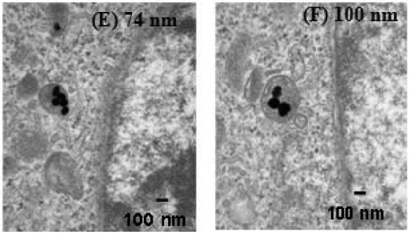
Fig 2 is an EM fig showing GNPs in endosomes, w/ some quantification: "Figure 2A shows that the number of nanoparticles per vesicle diameter is related to the size of the gold nanoparticles."
No information on the number of cells, vesicles, or nanoparticles analysed
No information on the number of cells, vesicles, or nanoparticles analysed
I find the error bars remarkably small, especially given the variability that one normally observe in such systems. And comparison with the images shown do not give me much confidence. 👇
For example. For the 100 nm GNPs, the authors report an average density of 1.6 10-3 GNP/nm2, but the one endosome shown in Fig 2F has 3 GNPs in an endosome of ~ 104,000 nm2, a density of 0.03 10-3 GNP/nm2.
For the 74 nm GNPs, the authors report an average density of 5 10-3 GNP/nm2, but one endosome shown in Fig 2E has 5 GNPs in an endosome of ~ 53,000 nm2, a density of 0.09 10-3 GNP/nm2. Another one has just one GNP for a similar sized endosome.
For reference, here is Fig 2A and Fig 2E-F. (Fig 2 also suffers from the same problem as Fig 1: it is unclear which concentrations were used; that would obv affect the results)

I have already mentioned Fig 3 in relation to effect of concentrations. Fig 3 also shows uptake as a function of time. Fine.

One interesting aspect to consider is that this kind of things had been done 50 years earlier and apparently forgotten. Here is Gosselin et al looking at the dynamics of uptake of gold nanoparticles in macrophages... in 1956.
ncbi.nlm.nih.gov/pmc/articles/P…
ncbi.nlm.nih.gov/pmc/articles/P…

and here is gold nanoparticles in endosomes in Hela cells... from 1957
ncbi.nlm.nih.gov/pmc/articles/P…
ncbi.nlm.nih.gov/pmc/articles/P…

(I really like that latter paper... it has 77 citations; the last 6 citations are from me)
Back to #16, Chitrani et al. Fig 4 shows non-specific adsorption of serum proteins on gold nanoparticles. OK. Fig 5 study the effect of shape, but there is very little data shown and the experiments also suffer from same issue as in Fig 1, i.e. what is concentration?
#17, Huang et al, Cancer cell imaging and photothermal therapy in the near-infrared region by using gold nanorods
dx.doi.org/10.1021/ja0572…
dx.doi.org/10.1021/ja0572…
From the same group as #11 (El Sayed). Very similar, except using gold nanorods instead of gold nanoparticles. Gold nanoparticles are included for comparison. The two papers are a little bit too similar in fact: image reuse between Fig3 of #11 and Fig 2 of #17


That paper (#17) has been cited 4,516 times according to Google Scholar.
unroll @threadreaderapp


