,
9 tweets,
6 min read
Read on Twitter
When someone is diagnosed with cancer, part of the tissue is removed and put into a solution that fixes it in time and place (called a fixative).
The tissue is then cut into very thin sections, put on a slide, and stained with dyes that help pathologists to visualize the tissue.
The tissue is then cut into very thin sections, put on a slide, and stained with dyes that help pathologists to visualize the tissue.
I took an amazing histology and pathology course when I was doing my PhD at @RutgersU in New Jersey USA. (Hi Dr. Reuhl!)
The first and most common stain is hematoxylin & eosin (H&E). Here are two pictures of normal skin and cancerous skin. Can you tell the difference?
The first and most common stain is hematoxylin & eosin (H&E). Here are two pictures of normal skin and cancerous skin. Can you tell the difference?
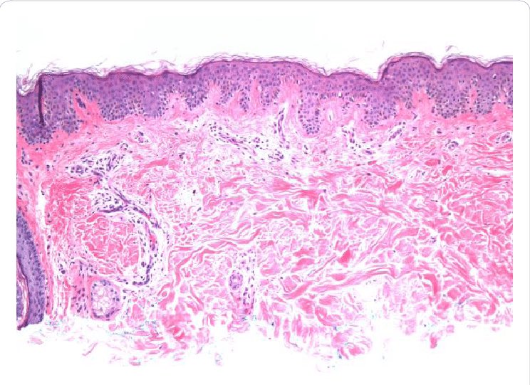
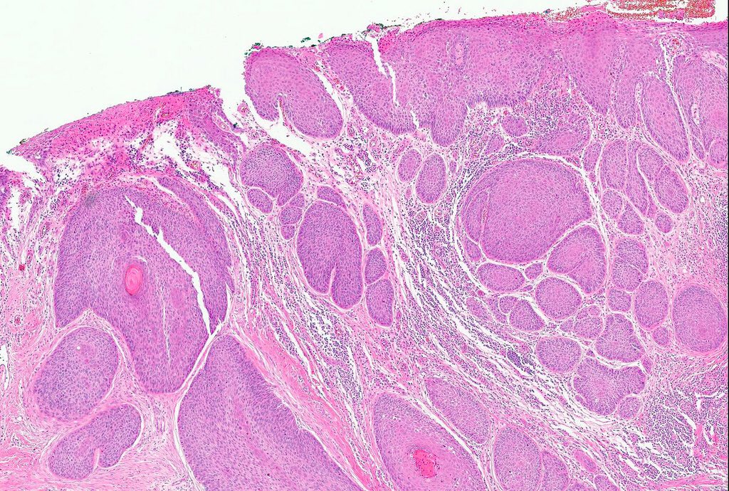
Hematoxylin stains nuclei dark blue or purple while eosin stains proteins in the cytoplasm of cells. While it’s great to show structure it doesn’t distinguish cells from connective tissue.
Enter: trichrome stain.
red= keratin and muscle fibers
blue= collagen
orange= RBC
Enter: trichrome stain.
red= keratin and muscle fibers
blue= collagen
orange= RBC

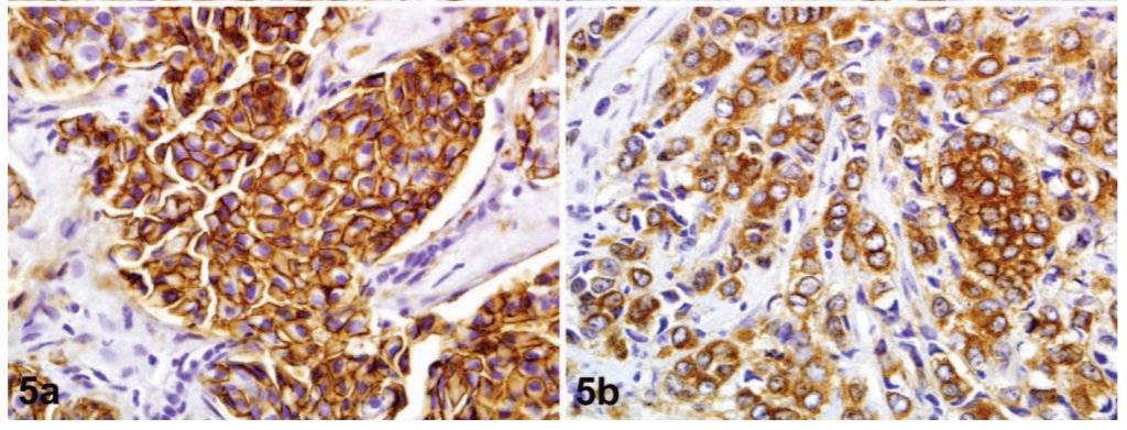
An important method is immunohistochemistry or IHC. This method uses antibodies to detect specific proteins in tissues.
In breast cancer, pathologists use this method to look at the estrogen receptor, progesterone receptor, and Her2. Brown = protein (More to come on these!)
In breast cancer, pathologists use this method to look at the estrogen receptor, progesterone receptor, and Her2. Brown = protein (More to come on these!)


One way pathologists determine the kind of breast cancer is based on histology or the way the cancer looks. The most type is ductal and the second most common is #lobular. (Hey @LobularResearch!)
#obular tumors lack the protein Ecadherin (Ecad) which is normally on the surface of cells attached to p120. Without Ecad, p120 is found in the cytoplasm (see picture from archivesofpathology.org/doi/pdf/10.585…; A= ductal, B= lobular):
#lobular is different from ductal in a few ways:
- it grows in a single file line (vs. as a round mass)
- it metastasizes to different sites in the body (ovary & peritoneum vs. liver & lungs)
- it responds differently to chemotherapy (more on treatments later)
- it grows in a single file line (vs. as a round mass)
- it metastasizes to different sites in the body (ovary & peritoneum vs. liver & lungs)
- it responds differently to chemotherapy (more on treatments later)
If you're interested in learning more about #lobular, follow @LobularResearch and @LobularBCA as well as research scientists @Prof_Riggins (my PI!), @mjsikora, and @oesterreichs. There's also a meeting focused on lobular breast cancer!

There are other histological subtypes of breast cancer as well including tubular, cribiform, medullary, and mucinous.