,
61 tweets,
22 min read
Read on Twitter
Did my fellows' curriculum lecture today, "Images in #Nephrology"
Thought of sharing the slides as a tweetorial. Goal is to present some 'classic' images that aid in kidney-related diagnoses. Not exhaustive; plan to make 2nd part as well.
#MedEd #FOAMed Not much POCUS involved!
Thought of sharing the slides as a tweetorial. Goal is to present some 'classic' images that aid in kidney-related diagnoses. Not exhaustive; plan to make 2nd part as well.
#MedEd #FOAMed Not much POCUS involved!


1st question. Classic image but rarely encountered. Note the super high blood urea nitrogen and serum creatinine.

Here is the answer.....

Another classic. It probably appeared on your USMLEs🤔
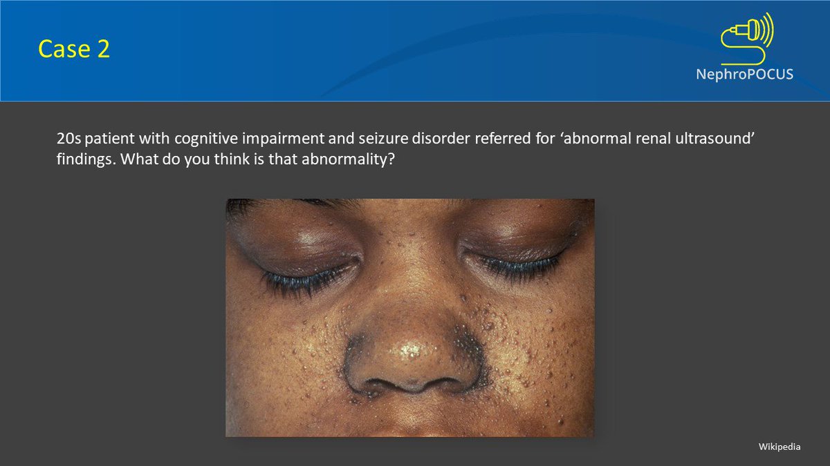
The diagnosis is Tuberous sclerosis complex (TSC) - genetic disease with autosomal dominant inheritance.
Clinical image shown: Facial angiofibromas
Kidney lesions to expect: Angiomyolipomas (AML) - most common followed by renal cystic disease.
Clinical image shown: Facial angiofibromas
Kidney lesions to expect: Angiomyolipomas (AML) - most common followed by renal cystic disease.

AMLs can be too numerous to count in TSC and in those cases, ultrasound is not good for surveillance. Obtain contrast MRI or CT.


Patients with TSC may be at increased risk for the development of renal cell carcinoma (RCC), most often the clear cell type. This is an example of RCC - note hypoechoic mass (unlike bright AML) and also calcifications.
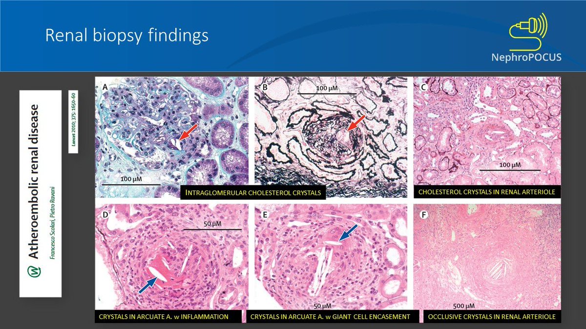
Some important things to know about these lesions.


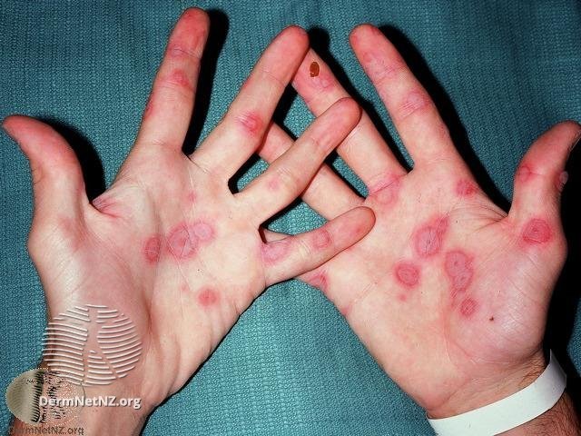
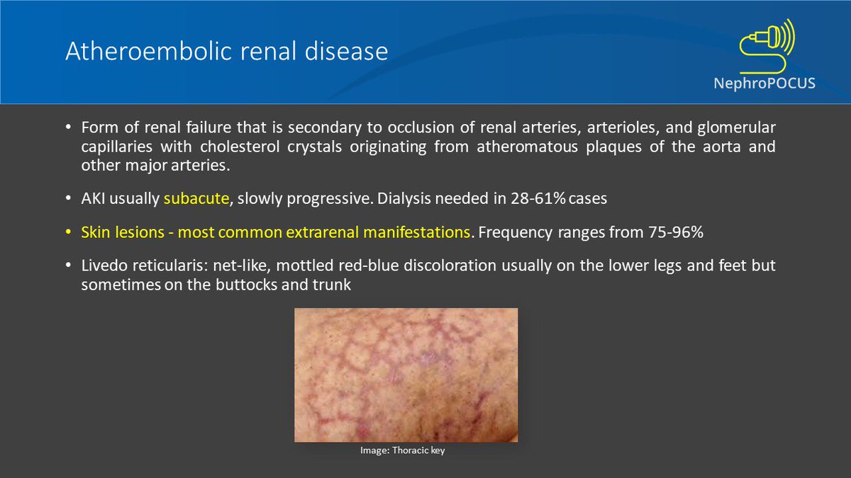
Here comes question 3 with classic skin lesions.

You are right...it's livedo reticularis.
Also note that the acute kidney injury is subacute and there is a delay between the precipitating event (typically PCI) and onset.
If you see AKI soon after PCI, it's probably contrast-induced or related to hemodynamic fluctuations.
Also note that the acute kidney injury is subacute and there is a delay between the precipitating event (typically PCI) and onset.
If you see AKI soon after PCI, it's probably contrast-induced or related to hemodynamic fluctuations.
Any laboratory clues to be aware of?

Very important images for the boards!
Must-know ophthalmoscopic finding in a patient with accelerated hypertension and headache/blurry vision.

Yes, it is papilledema. This images shows other findings in severe hypertensive retinopathy.

Do nephrologists go around carrying an ophthalmoscope?
Probably not. But they do carry echoscopes (at least some).
Don't be confused. Echoscope means ultrasound machine...it's a legitimate name!
Probably not. But they do carry echoscopes (at least some).
Don't be confused. Echoscope means ultrasound machine...it's a legitimate name!


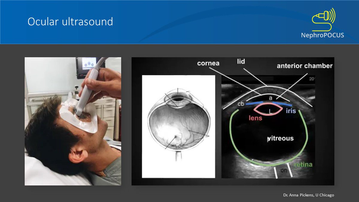
You are kidding me....how would you do #POCUS of the eye?
Here it is:
Use high frequency probe (vascular), put a lot of gel on the closed eyelid. Can apply transparent film dressing (Tegaderm) over the closed eye for patient comfort taking care not allowing air bubbles to form.
Here it is:
Use high frequency probe (vascular), put a lot of gel on the closed eyelid. Can apply transparent film dressing (Tegaderm) over the closed eye for patient comfort taking care not allowing air bubbles to form.
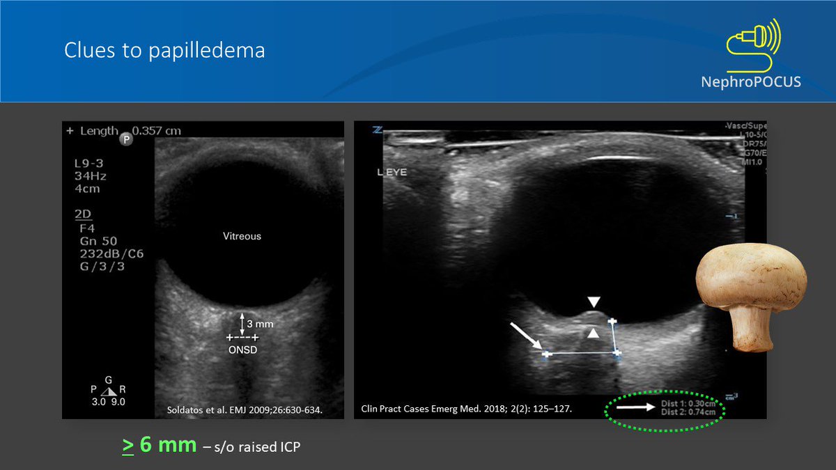
Measure optic nerve sheath diameter (ONSD) - hypoechoic stripe within the echogenic retrobulbar fat 3 mm posterior to the papilla. Upper limit of normal is considered to be ~5mm in general. Increases in papilledema. Also u may see optic disc bulging into vitreous like a mushroom.
Looks like some catheter-related infection. What about the eye? Should you prescribe some iv antibiotics with #Dialysis and leave it alone?
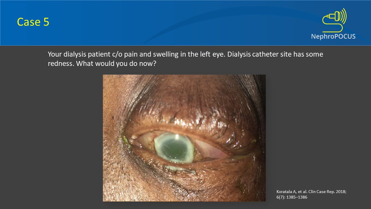
Don't wait...the eye doesn't look good. Obtain ophthalmology consult. . Endophthalmitis is a rare metastatic bacterial complication of dialysis access–related infection. Pic from my previous pub showing hypopyon.

Treatment is difficult because of poor penetration of systemic antibiotics into the vitreous humor, and early initiation of intravitreal antibiotic therapy plays a key role in the acute management of this potentially vision-threatening complication. Another case of mine 👇🏻

Here is a patient with advanced #CKD. What's your diagnosis?
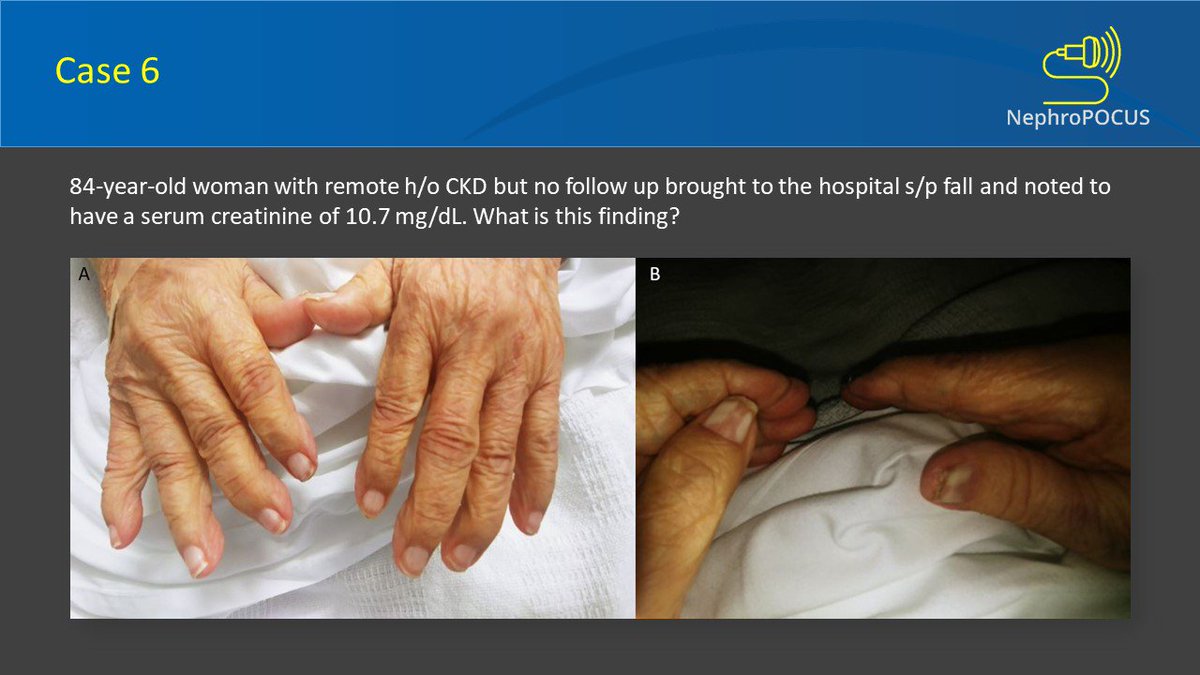
1/2 Answer: Half-and-half nails, also known as Lindsay’s nails - originally described as red, pink, or brown bands occupying 20 to 60% of the distal nail bed in patients with CKD. The proximal white band is thought to result from chronic anemia and the distal pink or brown
2/2 band from increased melanin deposition distally, possibly stimulated by uremic toxins. May be seen in conditions other than renal failure such as Crohn’sdisease (with or without associated zinc deficiency),Behcet’s disease and those on isoniazid therapy for TB.
Image: our own - Am J Med. 2019 Aug 14. pii: S0002-9343(19)30686-2. doi: 10.1016/j.amjmed.2019.07.042.
Doesn't look good 🤔
ESRD = End stage renal disease
STD = sexually transmitted diseases
ESRD = End stage renal disease
STD = sexually transmitted diseases


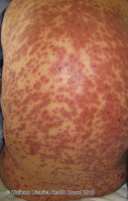
Saw the calcifications on plain X-ray?
That's suggestive of calciphylaxis (calcific uremic arteriolopathy is the preferred term)
Presents with skin ischemia and necrosis; characterized by calcification of arterioles and capillaries in the dermis and subcutaneous adipose tissue.
That's suggestive of calciphylaxis (calcific uremic arteriolopathy is the preferred term)
Presents with skin ischemia and necrosis; characterized by calcification of arterioles and capillaries in the dermis and subcutaneous adipose tissue.

This condition carries high mortality even with treatment.
Here are some images from my previous publications demonstrating vascular calcifications and corresponding lesions. Note that the calcifications can take various forms such as tram-track, patchy or linear.

Lanthanum (a phosphate binder used in CKD/ESRD patients) is a metal and can light up on X-rays. There is no evidence to suggest it causes harm to GI tract unlike another phosphate binder sevelamer.


Speak of the devil.....
I know you guessed it!
I know you guessed it!

The culprit is sevelamer, commonly used phosphate binder.

Here comes my #onconephrology favorite. Can you identify which 3 disease entities we are talking about?
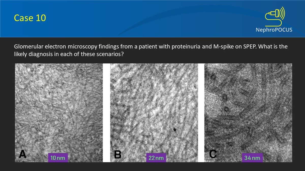
I'm sure you read about these hard-to-treat GNs but keep forgetting about the fibril diameter. Here is an easy way to remember: The size increases in alphabetical order - A, F, I and just remember 10-20-30 if not the ranges #nephpearls
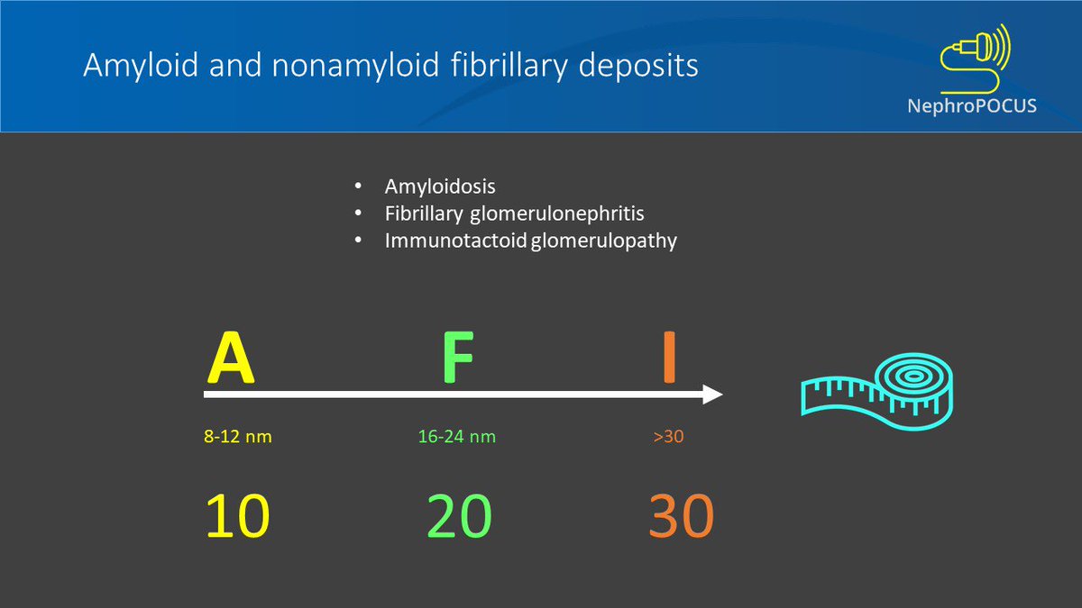
Of these, amyloid is the only one that stains positive for Congo red [at least for exam purposes].
Also, DNAJB9 is being much talked about these days. It has great sensitivity and specificity for fibrillary GN. Likely to appear on the boards.
Also, DNAJB9 is being much talked about these days. It has great sensitivity and specificity for fibrillary GN. Likely to appear on the boards.

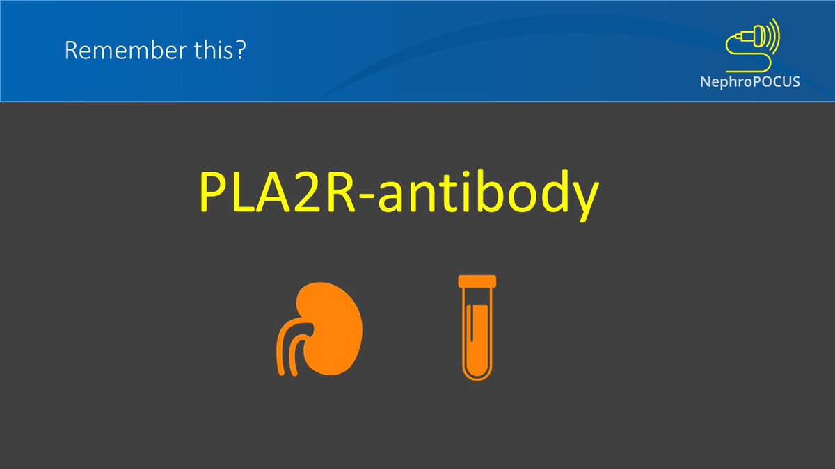
Another marker of practical significance in #nephrology is PLA2R. As you know, most (~80%) of 'primary' membranous GN are associated with auto-antibodies against podocyte antigen M-type phospholipase A2 receptor (that is, PLA2R). Can measure in blood also; used for monitoring.
Case number 11. This is another classic EM #biopsy image that appears on exams and quizzes.
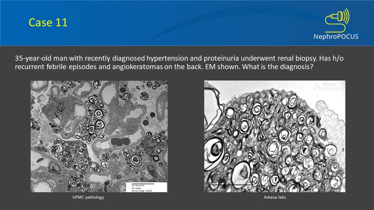
Need a clue?
Nothing better than this 🤓
Nothing better than this 🤓
Yess!! Zebra bodies.
ZEBRA = FABRY
X-linked: In general, males are more severely affected than females. Renal manifestations occur in at least 50% of male patients and ~20% of female patients.
May be an underdiagnosed disease.
ZEBRA = FABRY
X-linked: In general, males are more severely affected than females. Renal manifestations occur in at least 50% of male patients and ~20% of female patients.
May be an underdiagnosed disease.

Diagnosis: Initial - leukocyte alpha-galactosidase A (alpha-Gal A) activity.
Gold standard - Mutational analysis of the alpha-Gal A (galactosidase alpha [GLA]) gene.
Gold standard - Mutational analysis of the alpha-Gal A (galactosidase alpha [GLA]) gene.

So you saw Zebras on electron microscopy. Do you remember seeing something similar on light microscopy in another disease entity?
Clues: 1. Patients may present with symptoms mimicking malignancy; bladder and kidneys may be involved
2. Positive Von Kossa stain (stains calcium)
Clues: 1. Patients may present with symptoms mimicking malignancy; bladder and kidneys may be involved
2. Positive Von Kossa stain (stains calcium)

I know it's hard. The answer is Malakoplakia.
But important; this question appeared in KSAP (August 2019)
But important; this question appeared in KSAP (August 2019)

Michaelis–Gutmann bodies are concentrically layered basophilic inclusions within macrophages found in the urinary tract. Thought to represent remnants of phagosomes mineralized by iron and calcium deposits [The problem in Malakoplakia is defective phagocytosis]

Renal biopsy light microscopy trichrome stain in a #transplant patient on calcineurin inhibitor (Tacrolimus) therapy. Blue represents fibrosis on this stain.
Need to say more?
(Note: CKD stage 3T, T means transplant)
Need to say more?
(Note: CKD stage 3T, T means transplant)
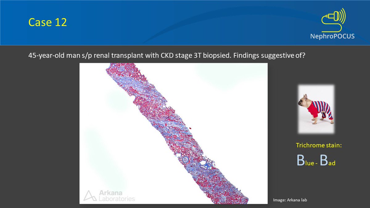
Yes, it is 'striped fibrosis' and represents chronic calcineurin inhibitor toxicity.

Here is a question on physical examination of the AV fistula.
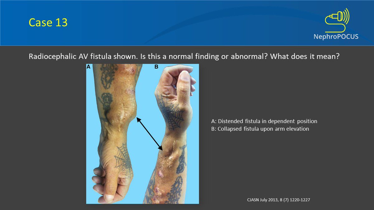
This is called the arm elevation test and abnormal test gives a clue to outflow stenosis.

This fistula is not good!
Further reading: This excellent tweetorial by @aishaikh
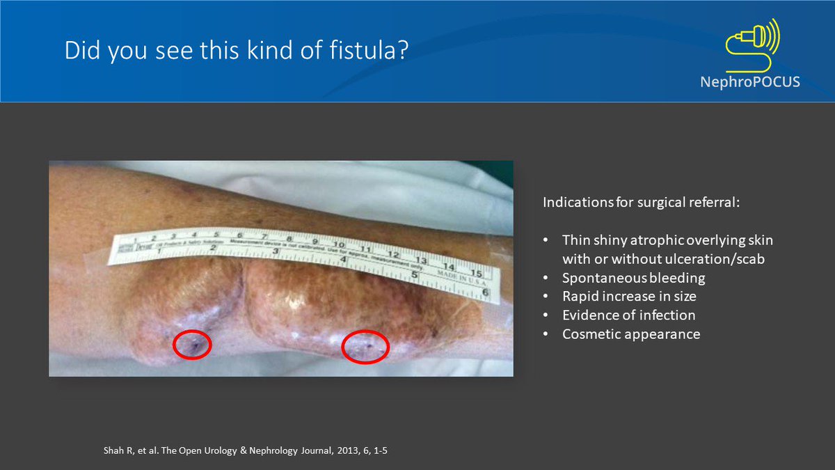
@aishaikh Another important scenario.
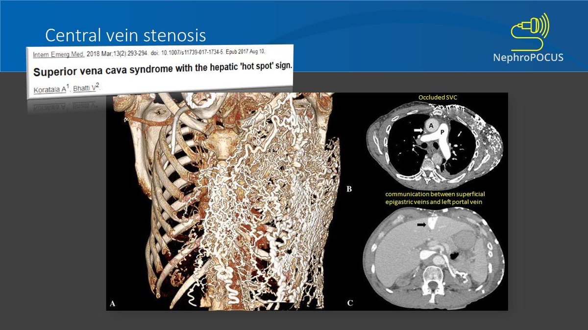
Beware of the central vein stenosis and refer to vascular surgeon appropriately.
Beware of the central vein stenosis and refer to vascular surgeon appropriately.
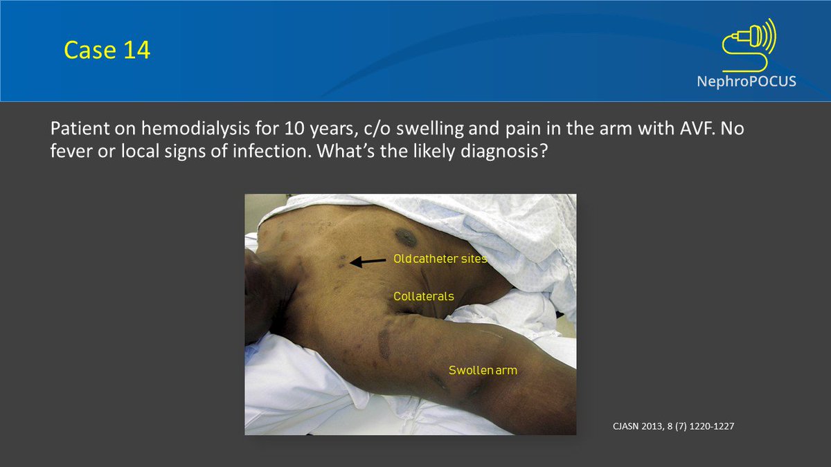
One of our old cases with extensive venous collaterals in a #dialysis patient with superior vena cava stenosis. CT shows near-complete occlusion of the vessel.
Last question of this #nephpearls tweetorial (for now).
Only one of the following rows accurately reflects the urinary crystal name and image. Which one is that?
Only one of the following rows accurately reflects the urinary crystal name and image. Which one is that?
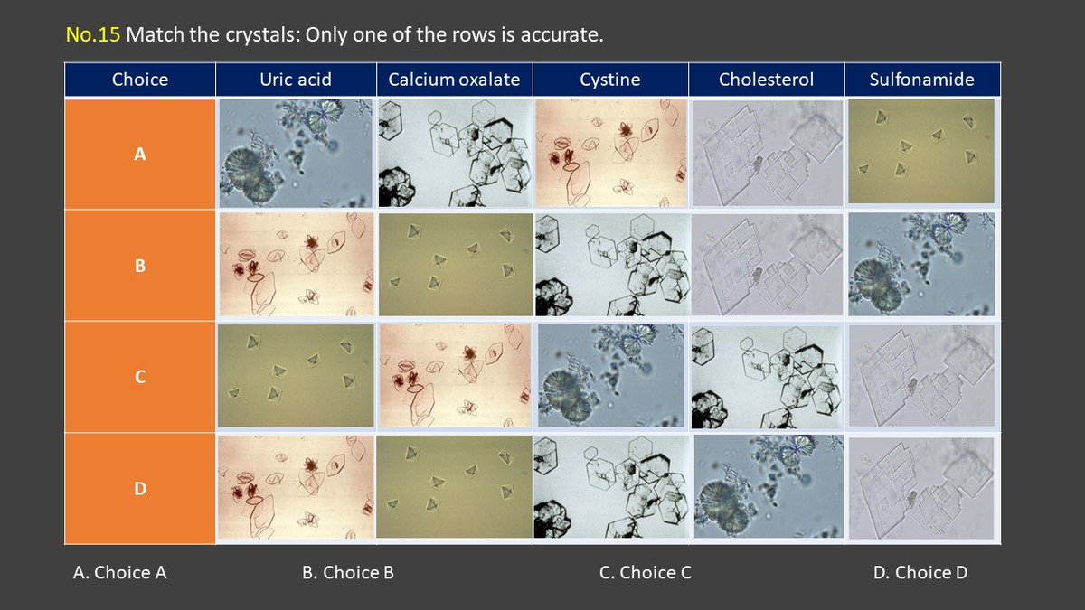
The answer is B.
Review the following slides quickly.
Review the following slides quickly.

Important scenario: Seen in ethylene glycol (anti-freeze) poisoning.
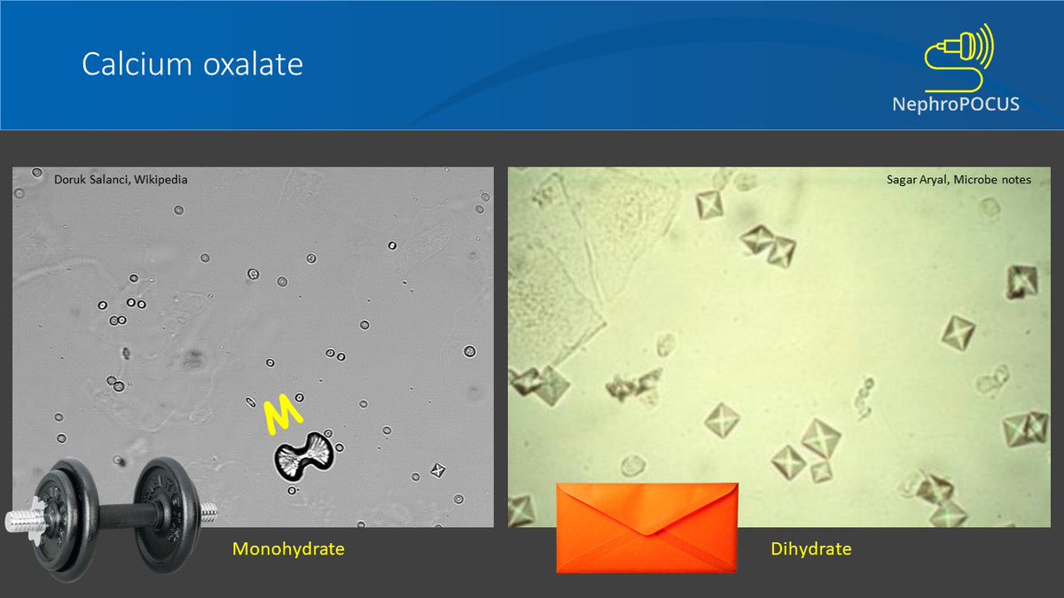
This shape is classic!

Frequently tested on board exam.

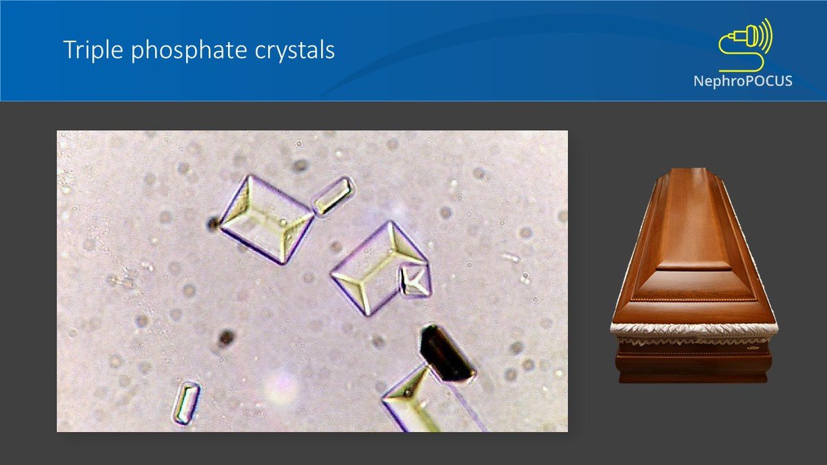
Tried hard to find the appropriate coffin lid 🧐
Suggested further reading: This excellent collection by @Neph_Sim
Done for now. Thanks for patiently going through all the 50+ slides (if you did 😉). Hope the tweetorial is of some use.
@kantsmd @RenalFellowNtwk @RegnerNephAKI @AmirKazory @UFNephrology
@kantsmd @RenalFellowNtwk @RegnerNephAKI @AmirKazory @UFNephrology

Only immunotactoid shows parallel arrangement. Rest of the two are irregular.