Happy 2021! @MBVanElzakker and I are excited to share our new article published in #Immunometabolism: “Pathogens Hijack Host Cell Metabolism: Intracellular Infection as a Driver of the Warburg Effect in Cancer and Other Chronic Inflammatory Conditions”: ij.hapres.com/htmls/IJ_1341_…
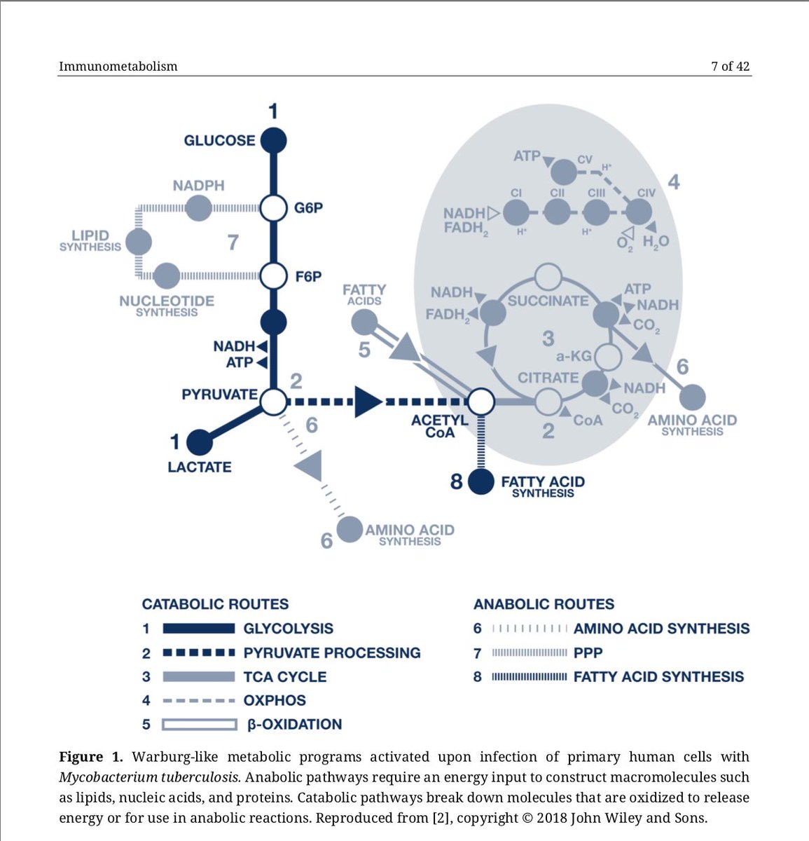
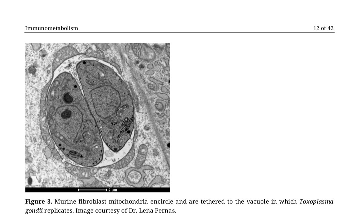
2/ In the paper, we detail molecular mechanisms by which #viral, #bacterial, and #parasite intracellular pathogens can induce, or contribute to, a Warburg-like #metabolism in infected host cells in order to meet their own replication and nutritional needs.
3/ We also discuss how host defense towards #infection may impact cellular metabolic changes (including how #mitochondria can participate in the innate immune response towards infection)
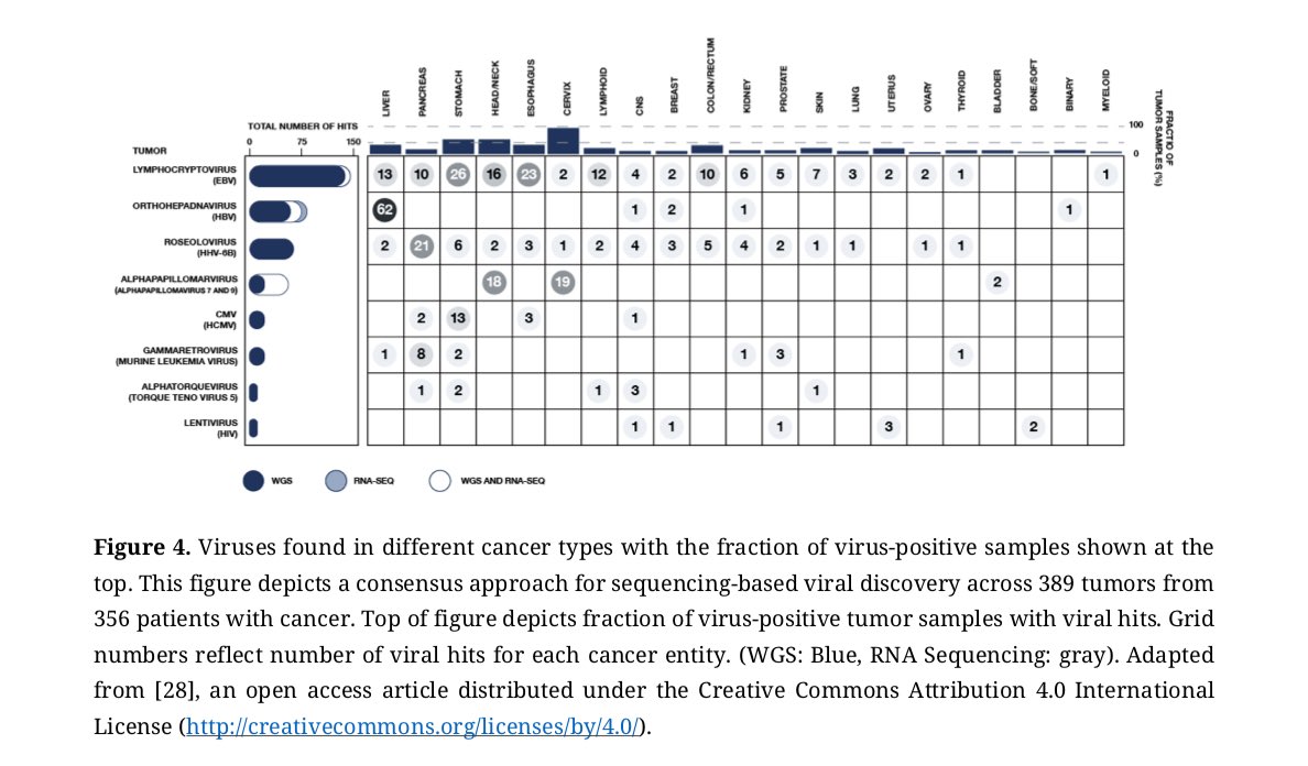
4/ We then provide examples of how many of these same intracellular pathogens have been identified in #tumors, atherosclerotic lesions, #granuloma, and other tissues containing cells with a Warburg or altered metabolism.
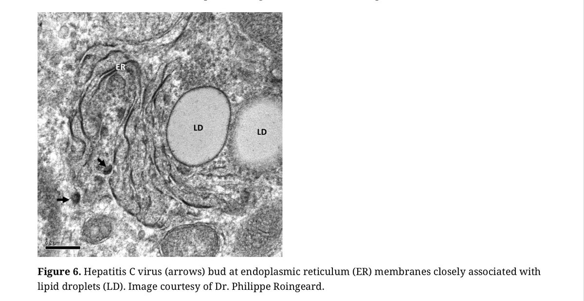
5/ Last, we examine further trends associated with infection and host cell metabolism, including how #pathogen-driven hijacking of host cell lipid metabolism can support viral, bacterial, and parasite survival and replication.
6/ Overall we argue that, at least in a subset of patients w/ #cancer, #atherosclerosis, #sarcoidosis + related conditions, metabolic changes and inflammatory cascades central to the disease process are driven by intracellular pathogens
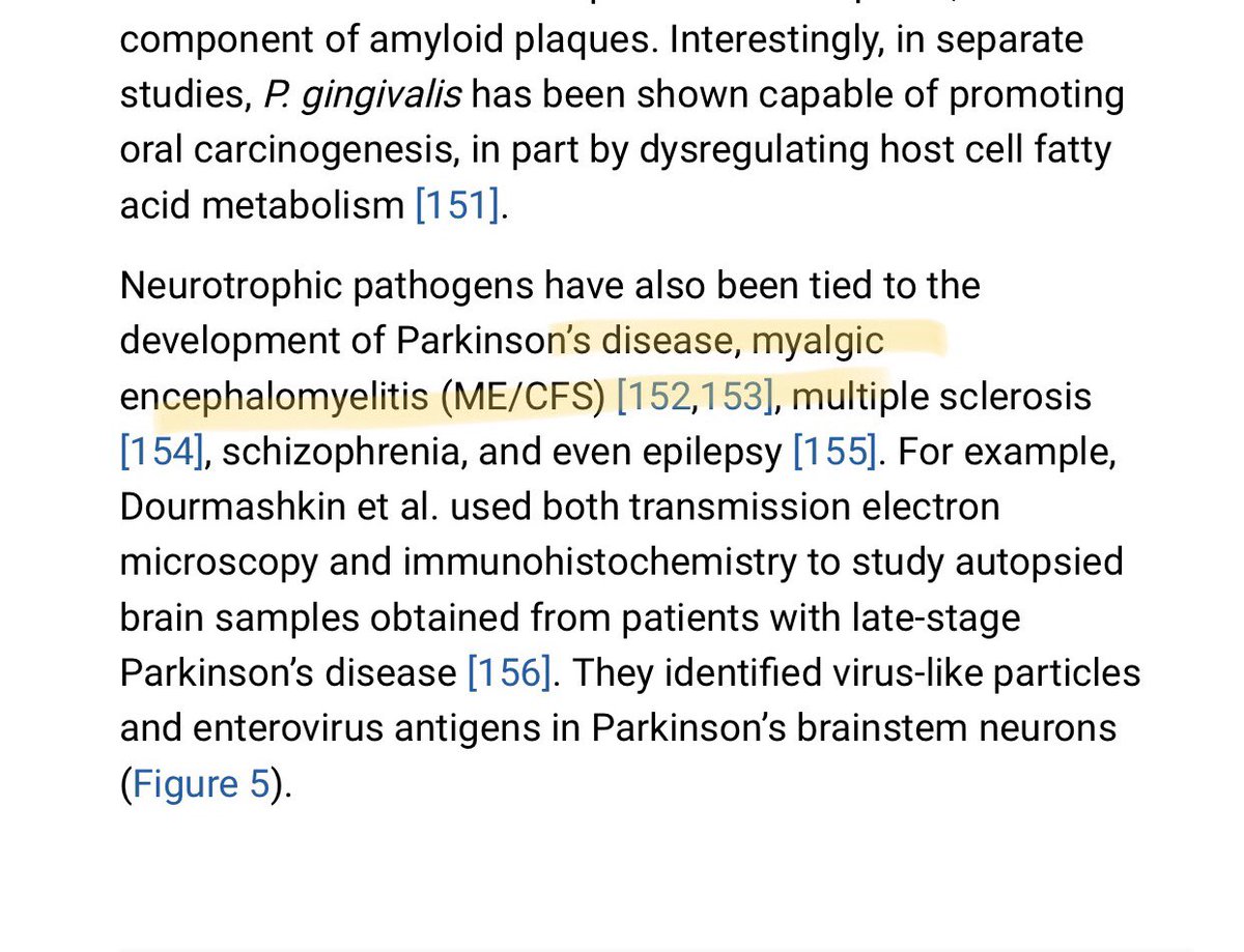
7/ We also discuss how pathogen-driven hijacking of cellular metabolism may contribute to #MECFS. ME/CFS is an extremely debilitating #neuroinflammatory condition that often begins with an infection (w/ #herpesvirus or enterovirus involvement already documented in some cases)
8/ A better understanding + acceptance of the trend could result in novel #diagnostics and treatments for patients w/ such conditions (w/ discussion of how a #ketogenic diet might impede pathogen-hijacking of host metabolism)
9/ So many cool people helped make the paper a reality. Special thanks to @lenapernas@Palmer_IMet_lab@dbkell, Dr. Wonder Drake, and @krisfobes for helping us with feedback and on earlier drafts of the manuscript.
• • •
Missing some Tweet in this thread? You can try to
force a refresh
It was inspiring to virtually attend the first Inaugural Robert D. Moir Symposium this past Friday. Rob was a friend and constant source of inspiration, plus an amazing sounding board for novel ideas. He passed away from glioblastoma one year ago today.
2/ Like most great scientists I’ve known, Rob was not content to study just the presence or absence of compounds/organisms in the human body. Instead his thinking continually gravitated towards the vital question of “what are they DOING??”
3/ Via that lens - the constant question of “why?” and a passion for characterizing the molecular biology of the “why?”...Rob uncovered that amyloid in the #Alzhiemer’s brain has a function (it appears to act as an antimicrobial peptide)
1/ This excellent study by @aaronmring/@VirusesImmunity and team found that COVID-19 patients exhibit dramatic increases in the production of antibodies against thousands of human extracellular and secreted proteins (the exoproteome) compared to controls 👇
2/ The million dollar question is: what molecular mechanisms underly this antibody/#autoantibody production? It is worth interpreting the findings via the lens of human #microbiome/#virome activity + the activity of persistent pathogens (such as EBV) harbored by study subjects.
3/ Every study subject harbored extensive microbiome/virome communities comprised of trillions of organisms during #COVID-19 infection…with such ecosystems now understood to persist beyond just the gut but also in other body sites (#lung, liver etc).
Important paper 👉 A team in Tokyo took RNA-seq data from the Genomic-#Tissue Expression Project: a public resource created to study tissue-specific gene expression/regulation from 54 tissue types collected from 1000+ healthy individuals at autopsy: bmcbiol.biomedcentral.com/articles/10.11…
2/ They successfully identified 39 viral species in at least one tissue (tissue types included #brain, pituitary, esophagus, thyroid, #heart, breast, lung, kidney, adrenal gland, prostate, #nerve, adipose tissue, blood vessel, ovary, uterus etc)
3/ Viruses identified in the various tissue samples included EBV, HSV-1, Varicella, CMV, HHV6-A/B, HHV-7, HCV, HPV, adeno-associated virus...and 16 RNA #viruses including Respiratory syncytial virus (RSV), Parainfluenza Virus 3..
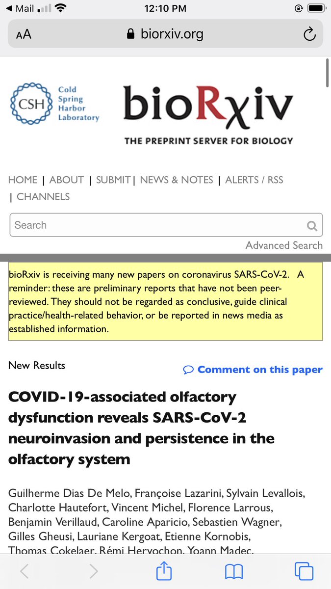
This is an incredibly important preprint to inform #LongCovid. Among many analyses, the team recruited 4 patients w/ prolonged + recurrent olfactory function loss after #COVID-19 (time from first COVID-19 symptoms to inclusion ranged from 110-196 days): biorxiv.org/content/10.110…
2/ None of these patients had detectable COVID-19 #RNA in nasopharyngeal samples by routine diagnosis (RT-qPCR). However, ALL patients had detectable COVID-19 RNA in samples obtained from their olfactory mucosa (confirmed with aRT-qPCR SYBR technique)
3/ Three of the patients had a high COVID-19 #viral load in the olfactory mucosa. Immunostaining additionally revealed the presence of COVID-19 antigens in 3 out of 4 patients. Based on that and related findings the team concluded...
@jenbrea Hey! So I'm getting to the point where I could write a long paper about potential overlapping connections b/t #Alzheimer's and #MECFS, but I'll summarize a few top trends in this thread! First, I brainstormed often on topic with the late Rob Moir
@jenbrea 2/ Rob’s research (done w/ Rudy Tanzi + Will Eimer at Harvard) forms the core of a potential ongoing paradigm shift in Alzheimer’s - namely that #amyloid beta may be an antimicrobial peptide that forms in response to pathogens in #brain tissue: pubmed.ncbi.nlm.nih.gov/30001512/
@jenbrea 3/ To better understand that research, read this #interview I did with Rob before he passed away last year from glioblastoma. Key to ME/CFS is his work indicates that amyloid may form in response to herpesviruses like HSV1 in the Alzheimer’s brain: microbeminded.com/2017/12/18/int…
This interesting study found that skeletal #muscle cells of #ME/CFS patients showed a decrease in oxidative phosphorylation (a metabolic pathway used by #mitochondria to generate #energy). In simple terms, that caused the cells to have a dysregulated #metabolism.
2/ Not mentioned in the paper, but important to consider, is that most well-studied #viral + bacterial #pathogens “hijack” the metabolism of the cells they infect in a manner that can result in decreased oxidative phosphorylation (or similar changes in cell energy pathways).
3/ In simples terms, these #pathogens “hijack” cell metabolism to “pull” substrates out of the human mitochondrial energy pathways...and use the substrates (lipids, fatty acids, amino acids) for their own #nutritional and replication purposes!