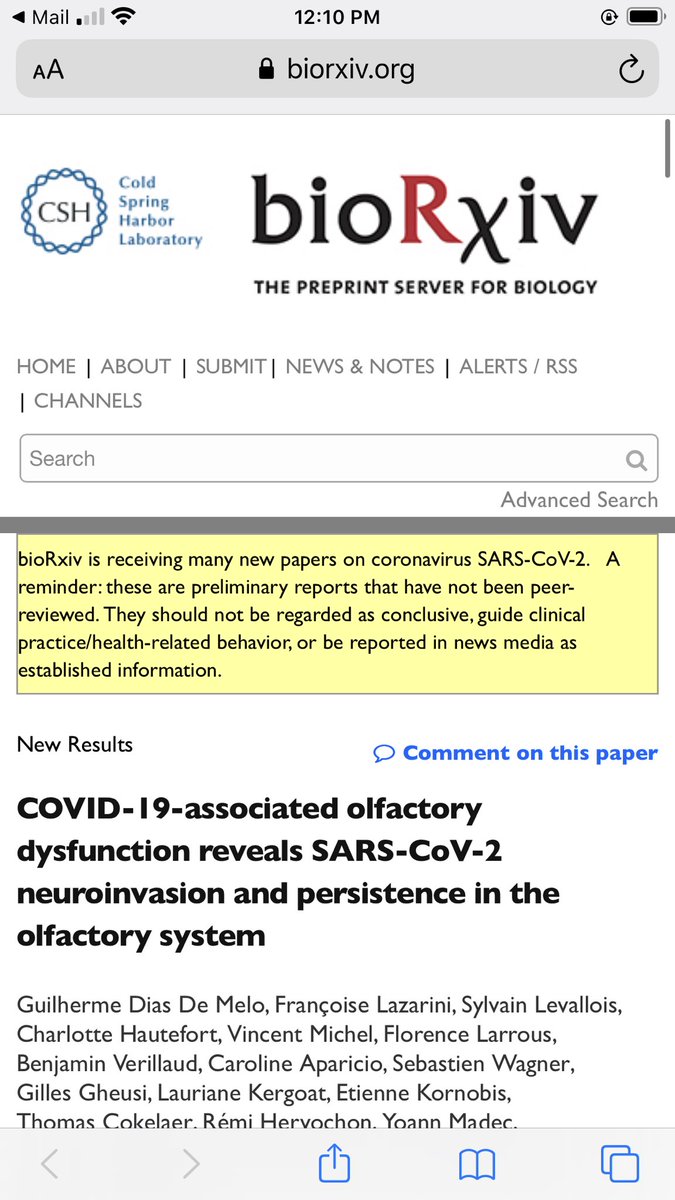
1/ This excellent study by @aaronmring/@VirusesImmunity and team found that COVID-19 patients exhibit dramatic increases in the production of antibodies against thousands of human extracellular and secreted proteins (the exoproteome) compared to controls 👇
https://twitter.com/virusesimmunity/status/1337574470878449665
2/ The million dollar question is: what molecular mechanisms underly this antibody/#autoantibody production? It is worth interpreting the findings via the lens of human #microbiome/#virome activity + the activity of persistent pathogens (such as EBV) harbored by study subjects.
3/ Every study subject harbored extensive microbiome/virome communities comprised of trillions of organisms during #COVID-19 infection…with such ecosystems now understood to persist beyond just the gut but also in other body sites (#lung, liver etc).
4/ Nearly all bacterial/viral/fungal members of such communities are #pathobionts: organisms capable of changing their gene expression to act as pathogens under conditions of imbalance/immunosuppression: ncbi.nlm.nih.gov/pmc/articles/P…
5/ This pathobiont activity can lead to increased expression of pathogenic proteins. And the human #immune system often responds to a pathobiont or its proteins by creating #antibodies capable of cross-reacting with human tissue/receptors etc via molecular mimicry.
6/ For example, this @Yale team identified pathobiont E. gallinarum in the mesenteric veins, mesenteric lymph nodes, liver, and spleens of mice made genetically prone to autoimmunity....: science.sciencemag.org/content/359/63… 

7/....In these mice, E. gallinarum initiated the production of “#autoantibodies,” activated T cells, and #inflammation. However this “autoantibody” production stopped when E. gallinarum’s growth was suppressed with the antibiotic vancomycin.
8/ ...Indeed , anti-dsDNA, anti-RNA autoantibodies, anti-b2GPI immunoglobulin G, hepatic and serum ERV gp70, and anti-ERV gp70 immune complexes were all suppressed by vancomycin treatment.
8/ Or consider this study: the team tested healthy women for the presence of IgG/IgA autoantibodies directed against 14 key regulatory peptides including leptin, ghrelin, vasopressin, and insulin..... pubmed.ncbi.nlm.nih.gov/18262391/ 

9/ ....Numerous cases of sequence homology were identified between these #peptides and the protein structures of over 30 #microbes including Listeria monocytogenes, E. coli, Lactobacilli, H. pylori etc.
10/ Or this study: B cells infected with Epstein Barr #Virus (EBV) secreted antibodies capable of reacting with dozens of self and non-self antigens including albumin, renin, and thyroglobulin: ashpublications.org/blood/article/… 

11/ So, is it possible that in #COVID-19 patients, at least some “autoantibody” production may be driven by changes in microbiome/virome pathobiont/#pathogen activity that occur when COVID-19 dysregulates the overall immune response?
12/ Certainly that would help explain the diversity of “autoantibody” responses identified in COVID-19 patients in the original @VirusesImmunity study (“autoantibody” production wld reflect activity of the unique microbiome/virome/pathogens harbored by each study subject).
13/ It would be interesting to create a #database of common human pathobionts/proteins…and screen the “autoantibodies” identified in the COVID-19 study against this microbiome/virome-derived #proteome/#metabolome for sequence homologies.
14/ For more context on the general molecular mechanisms underlying my thinking, please read my paper “Re-framing the Theory of #Autoimmunity in the Era of the Microbiome: Persistent Pathogens, Autoantibodies, and Molecular Mimicry”: discoverymedicine.com/Amy-D-Proal/20…
• • •
Missing some Tweet in this thread? You can try to
force a refresh


